News • Noninvasive stratification
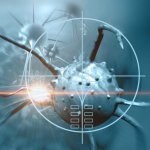
Multiplex PET helps doctors 'see' bowel cancer
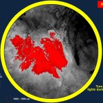
Multiplex PET imaging technology could provide a ground-breaking new approach for diagnosing and treating bowel cancer patients, according to scientists in Glasgow.


Multiplex PET imaging technology could provide a ground-breaking new approach for diagnosing and treating bowel cancer patients, according to scientists in Glasgow.
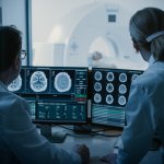
Radiologists and AI don’t always work well together: New research finds that the benefits of using AI tools appear to vary, boosting performance of some clinicians, but hurting others'.

Striking the balance between diagnostic efficacy and patient safety remains critical when utilising iodinated contrast media to deliver the best imaging outcomes. While playing a crucial role in diagnosis and treatment of disease, CT expert Efthimios Agadakos believes the medical profession has a duty to do its utmost to minimize patient risk from contrast media.
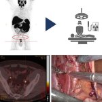
Dutch researchers use PSMA targeting to improve detection of prostate cancer, improving nodal staging and guiding more accurate surgery for this important patient population.
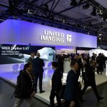
United Imaging showcased its full portfolio of AI-empowered products at the ECR 2024 in Vienna. The company highlighted its significant EU growth since establishing its regional HQ in 2019.
An international study has revealed that MRI monitoring in women with mutations in the BRCA1 genes significantly reduces breast cancer mortality without the need for preventive mastectomy.

As artificial intelligence (AI) is increasingly used in radiology, researchers caution that it’s essential to consider the environmental impact of AI tools.
Hologic, Inc. unveils new research in artificial intelligence (AI) and offering innovative educational opportunities at the annual European Congress of Radiology (ECR) in Vienna, Austria.
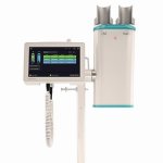
The Accutron CT-D Vision is the next generation of Medtron AG’s leading CT contrast media injector. Focusing on the user’s needs, the latest stage of development of the Accutron CT-D improves the usability of the CT injector and optimizes its integration into the radiological environment.

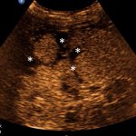
An innovative solution based on nanotechnology and ultrasound could prevent over-treatment of patients with rectal cancer. The magnetomotive ultrasound system uses nanotechnology for reliable diagnosis of any spread of rectal cancer to nearby lymph nodes.
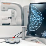
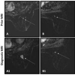
Ordering preoperative breast MRI exams of diagnosed breast cancer patients used to be controversial: Did they aid surgical planning better than the combination of mammography and breast ultrasound? Or did their findings cause overtreatment, specifically mastectomy, when breast-conservation surgery would have sufficed? New research has now settled the issue.
Stagnation, under-use, unfulfilled potential: At the EUSEM congress in Barcelona, leading emergency physician Dr Joseph Osterwalder describes how e-FAST (Extended Focused Assessment with Sonography for Trauma) – a key point-of-care ultrasound technique for trauma – has changed over the last two decades, and not necessarily for the better.

Breast cancer has no “one size fits all” therapy approach: subtypes differ significantly in malignancy, progression, and treatment response. Therefore, the more is known about the type of carcinoma in a patient, the better the outcome. At the annual scientific EUSOBI meeting in Valencia, Dr Ramona Woitek pointed out the potential of novel imaging techniques and computational image analysis…

‘Next Generation Radiology: Embracing the future and redefining the field of radiology’: In the run-up to the European Congress of Radiology 2024, we spoke with ESR President Professor Carlo Catalano from Rome, Italy, about the meeting’s content and its intriguing theme.
Using light instead of x-rays, a new imaging method from Philips is designed to advance navigation through a patient's blood vessels during minimally-ivasive procedures.
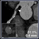
Higher image quality, better spatial resolution: A new radiology study demonstrates how photon-counting CT imaging can improve evaluation of coronary artery disease.
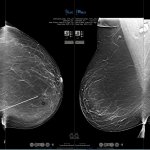
Breast MRI is increasingly being used as a primary breast cancer screening exam for young women. It brings benefits in women with dense breasts, who are at an elevated risk of developing breast cancer. The technique is also being ordered as a supplemental screening exam following mammography or breast ultrasound for women of all ages at high risk. But use of breast MRI as a screening tool is…
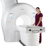
Fujifilm Healthcare Europe will present its Echelon Synergy MRI system at the European Congress of Radiology 2024. The 1.5 T scanner employs AI features to enhance image quality and scanning speed.

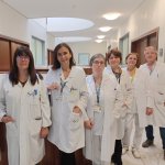
On 9 December 2023, NovaLife Polyclinic, with over 15 years of experience in the private healthcare sector in Timișoara, proudly inaugurated its state-of-the-art branch in the vibrant capital city of Bucharest.
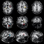
The loss of myelin sheaths in the brain is a hallmark of multiple sclerosis. Swiss researchers have now developed an MRI method that maps the condition of this nerve insulation layer more accurately.
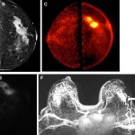
A new breast imaging technique provides high sensitivity for detecting cancer while significantly reducing the likelihood of false positive results, according to a new study.
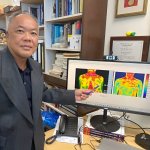
Researchers in Singapore have developed an AI-based software to assist in the early detection of breast cancer. Using thermal imaging, the program assesses the malignancy of a tumour.
Breast surgery is a traumatic experience for a woman, no matter whether breast-conservation surgery (BCS) or a mastectomy. Trauma levels are greatly enhanced, if pathological evaluation findings of an excised breast tumour following a lumpectomy suggest that additional cancer may still be in the margins, and a second surgical procedure is required. A new system with the ability to accurately…

United Imaging Healthcare Europe, a leading company in advanced medical imaging and radiotherapy equipment, proudly announces the introduction of its first Mobile Digital PET/CT solution in Italy, now fully operational in the Piacenza province under the auspices of the Azienda Unità Sanitaria Locale Piacenza (AUSLP) hospital.
RTI presents its latest advancement in X-ray Quality Assurance and testing. Centered around efficiency, Mako provides the ultimate no-fuss experience, simple setup in all X-ray applications, and fully wireless capability.