Image source: Queensland Brain Institute
News • Understanding the mechanisms
New ultrafast fMRI technique to improve brain stimulation
Brain stimulation, such as deep brain stimulation (DBS), is a powerful way to treat neurological and psychiatric disorders. While it has provided therapeutic benefit for sufferers of Parkinson’s, Alzheimer’s, and addiction for more than a decade, its underlying neural mechanism is not yet fully understood.
The study was first published in the Proceedings of the National Academy of Sciences (PNAS).
Researchers at the Queensland Brain Institute (QBI) are now one step closer to unravelling the mystery of brain activity to better understand this mechanism and potentially predict DBS outcomes.
The brain is a highly complex network of circuits organised hierarchically with wide-ranging connections. Connections go in different directions, forwards and backwards, and between neurons that are either excitatory – the accelerators of a response – or inhibitory – the brakes modifying a response. “Say you see an apple and want to grab it – once that sensory signal reaches the brain, we expect that the activity that follows depends on the brain’s neural connections,” Associate Professor Kai-Hsiang Chuang said. “What we don’t fully understand is how or when these structural and functional components of the brain process and integrate information to eventually lead to the outcome of moving your hand.”
Brain activity not only propagates through structural wiring but follows certain preferential circuits depending on their excitatory and inhibitory neuronal distribution
Kai-Hsiang Chuang
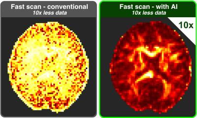
Functional MRI (fMRI) is the most popular technique used to study brain networks. fMRI tracks blood flow and oxygenation changes following neural activity, thereby indirectly measuring the functional connections being formed, and giving us an indication of where brain activity is propagating. Brain activity, however, isn’t as simple as signal travelling from area to area.
The team at the Chuang laboratory have developed a new ultrafast fMRI technique with vastly increased temporal resolutions, enabling them to capture the dynamics of brain activity at a sub-second level. Associate Professor Chuang said the new technique had led to more comprehensive understanding of how and when the brain’s structural and functional connections interact. “The first new discovery we made is that brain activity not only propagates through structural wiring but follows certain preferential circuits depending on their excitatory and inhibitory neuronal distribution,” he said. “Communication between brain regions of similar cell types becomes more fluent, and the brain activity stronger.”
The Chuang group tracked the brain activity of mice both while stimulated and at rest using their ultrafast fMRI technique. When the brain was stimulated, activity followed the structural wiring in the forward direction — from A to B and then B to C. When the brain was at rest, activity was more dependent on cell type organisation and less rely on this structural wiring, propagating between C and B but not with A, if that’s where the preferential circuit was. That means how information is processed is actually dependent on your state, where it was previously thought that brain activity functioned in the same way whether at rest or busy doing a task. “The second discovery we made was that the blood signal detected by fMRI could reflect the network organization and cell type distribution,” Associate Professor Chuang said. “These findings have significant implications for how brain structure shapes function, and how to predict activity based on the knowledge of this structure. More practically, what we now know will impact the design of DBS or other brain stimulation techniques. The next steps are to work with clinicians versed in brain stimulation to determine how we can utilise this knowledge combined with human data to help improve our understanding of DBS.”
This more comprehensive understanding could enable us to better predict DBS results and potentially improve its design for better therapeutic outcomes.
Source: Queensland Brain Institute
30.01.2023