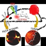
Imaging glucose uptake inside single cells
Researchers at Columbia University have reported a new approach to visualize glucose uptake activity in single living cells by light microscopy with minimum disturbance.



Researchers at Columbia University have reported a new approach to visualize glucose uptake activity in single living cells by light microscopy with minimum disturbance.
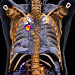
Dr Gerald Antoch, professor of radiology and chairman of the department of diagnostic and interventional radiology at Düsseldorf University Hospital and active member of several scientific societies, delivered the prestigious Wilhelm Conrad Röntgen Honorary Lecture at ECR 2015 on ‘Hybrid imaging: Let the two worlds of radiology and nuclear medicine come together’. Report: Marcel Rasch

The importance of cardiac imaging is increasing, but nuclear medicine procedures are by no means obsolete, observes Okan Ekinci, during our EH interview with the Siemens Vice President for Healthcare Consulting & Clinical Affairs about the latest innovations for cardiology.
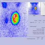
Before 2013, when Professor Dietmar Dinter became partner of Radiologie Schwetzingen, a multi-discipline group practice specialised in radiology and nuclear medicine, he was senior resident at the Institute of Clinical Radiology and Nuclear Medicine at University Hospital Mannheim (2003-2012) and head of its Nuclear Medicine Department (2009-2012). Was his work in nuclear medicine altered by the…

What's the ideal solution for vulnerable plaque imaging? 'A non-invasive imaging procedure with high spatial and temporal resolution, and without radiation exposure, and which provides information on coronary plaque composition precisely and in series.' Report: Axel Viola
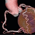
The significant benefits of cardiac catherisation remain undisputed. However, cross-sectional imaging modalities are serious competitors when it comes to arriving at the right diagnosis.

A session at the forthcoming British Cardiovascular Society annual conference (2-4 June, in Manchester) will hear about the latest imaging techniques for acquired heart disease, with PET technology playing a key role in those advances.

Claron Technology, the leader in universal medical imaging viewers, announces that it has been awarded the CE mark for its NilRead zero-footprint viewer for the diagnostic interpretation of medical images.
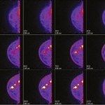
US-American researchers have shown that positron emission tomography (PET), a tried procedure, is a helpful modality to detect breast cancer.
However, investments in equipment and advanced training are attracting medical students, John Brosky reports
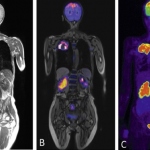
European Hospital met up with Professor Harald H. Quick, PhD, who was appointed Director of the Erwin L. Hahn Institute (ELH) for Magnetic Resonance (MR) Imaging this February.

The multiple benefits of PET/CT are undisputed – one being the fact that radiopharmaceuticals, which are used at pico and nano levels – are not toxic.

Screening for lung cancer saves lives. This fact has been documented by outcomes of the U.S. National Lung Screening Trial (NLST) which showed a 20% reduction in lung cancer-specific deaths in patients who had a chest CT screening. What is controversial is how to establish the radiology resources needed to perform exams for all the people who need it and then how to pay for the exams.
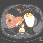
With MRI and CT scanners widely available in clinical routine, radiologists cull increasingly precise and relevant functional tumour information for diagnostics and monitoring purposes. Both modalities offer technological and methodological approaches, initiated by the discipline itself, that have become indispensable for certain frequent tumours.

The potential of cardiac magnetic resonance imaging (CMRI) is still largely untapped. One novel application might be ablation follow-up. The first MRI-guided cardiac interventions were performed at Herzzentrum Leipzig, but, as far as coronary imaging is concerned, MDCT remains superior to MRI

How the different advanced cardio vascular imaging technologies fit together in managing cardiac patients, will be one of the main themes explored at the International Conference on Nuclear Cardiology and Cardiac CT (ICNC 11).

Staging of cervical cancer is clinically based on a system developed by the International Federation of Gynaecology and Obstetrics (FIGO). The staging is clinical because the majority of cases occur in developing countries where access to MRI is limited, explains gynaecological radiologist Dr Evis Sala.
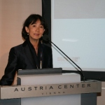
For the 19th consecutive year the annual meeting of the European Society of Radiology (ESR), the European Congress of Radiology (ECR), takes place in Vienna.
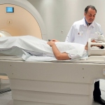
Experts across Europe believe the combination is beginning to demonstrate its broad potential as a hybrid imaging tool

The last decade saw PET and now PET/CT numbers expand rapidly in Europe. More recently the picture has been less rosy with public and private sectors delaying new projects and pushing back the replacement of equipment.


Dr Peter Choyke, Chief of the Molecular Imaging Programme at the National Cancer Institute in Bethesda, USA, believes that new tracers will have an evolving role to play and represent an exciting development in the imaging of cancers.

When asked about his vision of imaging in the year 2020, Professor Bernd Hamm MD, director of the three radiology clinics at the Charité University Hospital in Berlin, qualified his focus: ‘Technology is always only a vehicle. When we talk about road traffic, we don’t talk about the design of cars but about structural issues’

Virtual FDG-PET/CT bronchoscopy has been found to be a technically feasible tool for the detection of lymph node metastases in non-small cell lung cancer patients with good diagnostic accuracy, according to researchers at the Department of Diagnostic and Interventional Radiology, University Hospital Dusseldorf and Essen.

Quick, simple and comfortable – the RADbook 2012 on your iPad. Download the RADbook app free of charge today and have all state-of-the-art diagnostic imaging systems at your fingertips.

