Emergence of hybrid PET-MRI: first clinical experience
15:30–16:45
Session 2
Concerns: What can be done about the lack of young radiologists in Europe?
Prof. Osman Ratib, Head of the Department of Radiology and Nuclear Medicine, University Hospital; Geneva/CH
Whole-body hybrid PET-MR scanners are emerging on the market and are expected to have a significant impact in diagnostic imaging particularly in oncology applications and also in other clinical domains, such as cardiology, inflammatory and infectious disease, as well as in neurological applications.
He later then became Professor and Vice Chairman of the Department of Radiology at UCLA, and coordinated the deployment of an enterprise-wide strategy and infrastructure for image management and communication and seven years later, back at the University Hospital of Geneva, was made responsible for new molecular and functional imaging techniques and particularly of hybrid PET-CT.
His clinical activities include cardiovascular MR and CT imaging procedures, combined PET-CT imaging and advanced cardiovascular imaging. Today, as Chairman of the Department of Radiology and Head of the Division of Nuclear Medicine at the same university, he is responsible for six clinical divisions including radiology, neuroradiology, radio-oncology, nuclear medicine and medical informatics as well as a cyclotron and pre-clinical imaging unit. He has pioneered several innovative projects including advanced cardiovascular PET-CT programme and the first whole-body PET-MRI unit in Europe.
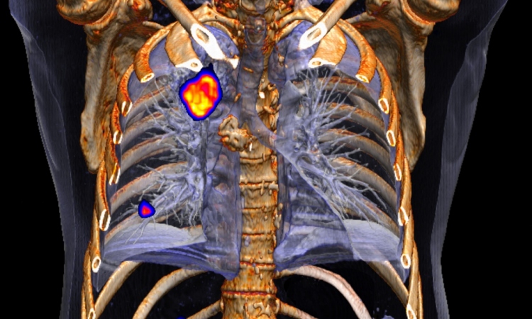
A whole-body hybrid PET-MR imaging unit was implemented at the University Hospital of Geneva and tested against PET-CT imaging for diagnostic and follow up of oncology patients. The whole-body PET-MR scanner, consisting of a 3-T MR and a time-of-flight PET scanner sharing a single bed allowing sequential acquisition of co-registered MR and PET images, was evaluated clinically in patients referred for diagnostic PET-CT study. PET-MR images were acquired following standard clinical PET-MR studies. Oncological studies included lymphomas, head and neck tumours, prostate and breast tumours as well as lung and colon cancers.
Optimised imaging protocols combining whole body MR attenuation correction data set with standard MR diagnostic protocols of both modalities while reducing the total time of the study were developed.
The diagnostic quality of fused PET-MR images was comparable to corresponding PET-CT images and measured local SUV were comparable. Advantages and limitations of this new technique compared to results obtained from conventional PET-CT technique will be reported.
11.02.2011