In their infancy but new PET tracers have a rich future
Dr Peter Choyke, Chief of the Molecular Imaging Programme at the National Cancer Institute in Bethesda, USA, believes that new tracers will have an evolving role to play and represent an exciting development in the imaging of cancers.

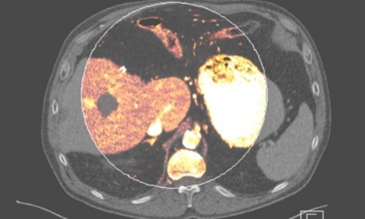
He will outline the potential of new PET tracers in a session at the ECR 2013 congress in Vienna next March. In an interview with European Hospital he explained why he believes they represent such an advance. Due to the sensitivity of PET (nanopico molar sensitivity vs. micromolar for MRI) it is possible, he said, to image cell membrane based receptors responsible for the abnormal growth associated with cancers and detect subtle changes in the integrity of cancer cells. FDG-PET/CT has been the trailblazer agent, demonstrating unique sensitivity for cancers, but he said while FDG uptake does reflect glycolysis, it is relatively non-specific and, to date, has not dictated the choice of therapies.
‘The promise of new PET agents is that they will aid clinicians in adding or deleting therapies depending on the pharmacodynamics of the imaging biomarker,’ he added. ‘For instance, classes of agents have been developed to investigate angiogenesis, proliferation, hypoxia, apoptosis, hormone sensitivity and amino acid transport. Each of these provides a unique window on the biology of each cancer and will hopefully guide therapies in the near future.’ In the specific example of metastatic prostate cancers, he said, Sodium Fluoride PET is proving far more sensitive than conventional bone scans. Agents such as F-ACBC (amino acid transport) F-DCFBC (Prostate Membrane Specific Antigen PSMA) F-DHT (androgen receptor) and F-Choline (cell membrane turnover) are proving efficacious in the detection of metastatic disease and reflect actual tumour burden in contrast to existing methods that only indirectly image tumour (bone uptake).
The new tracers he will mainly focus on at ECR 2013 will be F-18- FLT, F-18 Fluciclitide, F-18 FACBC, F-18 DCFBC, F-18 Estradiol and F-18 Sodium Fluoride - newer agents that are more specific and target receptors on cancer cells – and they have a clear application for oncology with F-18 Fluciclitide being useful in identifying tumours with high integrin expression or angiogenesis, possibly suggesting that an anti-angiogenic agent or anti-integrin agent would be a useful adjunct to the treatment.
‘Similarly,’ he added, ‘F-18 Estradiol (FES) shows whether the oestrogen receptor is active in a tumour. This can be useful in determining which drugs targeting ER are appropriate for a specific tumour.’ Some of the agents may also be useful in determining whether inflammation is present (e.g. in inflammatory bowel disease) or if there is ischemia (vascular disease). The major benefit of the newer tracers to clinicians/radiologists, he pointed out, will be in selecting the right drug for the right patient.
‘Currently, oncologists use the same kinds of drugs in all patients for first and second line therapies. Using new tracers it will be possible to select the best drugs for a patient and to anticipate when drug resistance is beginning, thus providing an opportunity to switch to more effective therapies before subjecting the patient to needless toxicities. Moreover, these tracers will allow earlier assessment of the benefit of particular therapies. Ineffective drugs can be dropped earlier while more effective drugs can be added to the regimen.’
For the patient, he pointed out, molecular imaging provides the opportunity to treat each tumour individually and monitor for success or failure by using specific markers expressed by the tumour, and be accomplished without invasive biopsies. They also may change the approach to diagnosis and treatment. ‘Molecular imaging, along with new tissue and serum/urine based biomarkers,’ Dr Choyke confirmed, ‘offer the possibility of customising treatments based on individual tumour molecular expression profiles.’
PROFILE
Dr Peter Choyke, Chief of the Molecular Imaging Programme of the National Cancer Institute, Bethesda, graduated from Jefferson Medical College and trained in Diagnostic Radiology at Yale University and the University of Pennsylvania. His research focuses on the development of new imaging methods, principally PET, MRI and optical, that result in improved outcomes for cancer patients. His special interest is prostate cancer for which he has an active research programme involving MRI, PET and optical imaging.
02.11.2012