CT and PET - Improving radiation therapy planning
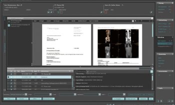
When planning radiotherapy the combination of positron emission tomography (PET) and Computed tomography (CT) can provide a better outcome than CT alone. Michael Krassnitzer asked Terri Bresenham MSc BSc, Vice President for Molecular Imaging at GE Healthcare, for her views on the value of PET/CT, the new EANM guidelines, novel tracers and the future of other hybrid imaging technologies.

Radiation therapy planning uses the anatomical structure to decide where best to focus radiation. ‘By heightening the information of the metabolic clusters it’s possible to spare more of the normal tissue and to deliver more intense radiation in the areas where there is more metabolic activity. Thereby, radiation therapy planning takes a leap forward. But we don’t forget to do some research on different tracers, in addition to today’s use of glucose,’ Terri Bresenham explained. ‘It’s a very exiting area, because the process of understanding cancer is very complex.’
The European Association of Nuclear medicine (EANM) recently published guidelines concerning the use of PET in radiotherapy planning. ‘Previously, there were only guidelines in some countries as well as for certain cancers,’ she pointed out. ‘The newly established guidelines are a pioneering work. For therapy planning of particular cancers – tumours in the brain, head and neck, lung, and also gastro-intestinal, genito-urinary and gynaecological tumours – the guidelines show that the outcome is better with the use of PET information. A lot of technology inherently has the disadvantage that scientific comparative effectiveness work has not been done to show the evidence. Doing this scientific work is a part of our responsibility.’
This why GE healthcare supported the working out of the guidelines with research funds, she pointed out. ‘In terms of technology innovation, GE Healthcare has a responsibility. We don’t look only at the technology; we try to look at improvements in quality and in the costs of care because, in the end, the long-term sustainability of healthcare technology relies on the fact that it brings a limitation of costs and demonstrated quality improvement.
New tracers
‘We are entering a period of discovery, in which everything that we know clinically today will completely change. There will be,’ she forecast, ‘a menu of specific tracers that we will use to get specific answers to our questions. You can couple this with advances in understanding the genomics, proteomics and metabolism. Knowing more about the basis of biology and biochemistry will enable us to build tracers that can identify those receptors or discover that biological process.’
Could CT remain the gold standard in radiotherapy planning? ‘CT is a wonderful tool. I don’t see that going away anytime soon,’ she believes. ‘Certainly the future of radiation therapy planning is CT accompanied by metabolic information from PET.’
What about other hybrid imaging techniques, e.g. PET/MRI and SPECT/CT (single photon emission computed tomography)? ‘What will be overtaken is the use of SPECT without any CT. In the user’s community there are many cameras that only have SPECT, hence no CT. That population of cameras gradually will be replaced with PET/CT, because they offer better diagnostics.
‘PET/MR is a very interesting idea. There are plenty of papers that say that PET/MR would be absolutely the right tool to use. However, for today -- and probably for the next five to ten years -- the PET/MR-combination will be applied more in laboratory research than in clinical use.’
03.11.2010
- CT (605)
- molecular (205)
- nuclear medicine (127)
- PET/CT (185)
- radiation therapy (180)
- SPECT (31)
- therapy (845)


