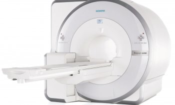
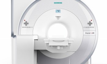
Hybrid imaging: PET/MR climbs the diagnostic ladder
Experts across Europe believe the combination is beginning to demonstrate its broad potential as a hybrid imaging tool

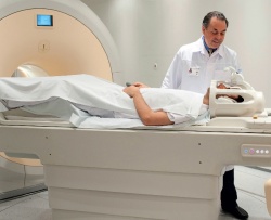
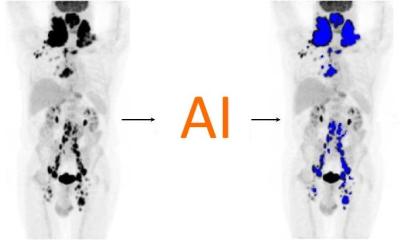
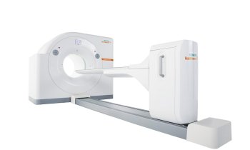
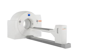
Whilst PET/CT remains the gold standard in hybrid imaging at this stage, PET/MR has shown great promise for imaging of head and neck cancers, prostate and breast imaging and, over the last two years, radiologists have recognised its value and potential as a diagnostic tool. However, evolution has not been pain-free for those pioneering its development. During the PET/MR: A marriage made in heaven or hell? New Horizons session (ECR 2012, 9 March. NH8. 8-10 a.m.) when the latest position on PET/MR will be presented, it will also serve to underline only too well that the journey has not been easy. Professor Osman Ratib, Chairman of the Department of Medical Imaging and Information Sciences and Head of the Division of Nuclear Medicine at the University Hospital of Geneva, Switzerland, is among the speakers. His department was the first in Europe (2010) to have whole body PET/MR. With questions over whether such technology will replace, or complement, PET/CT, he said that the session would offer a clinical perspective on this new hybrid modality.
‘We know there are problems; that’s why we have the ‘heaven and hell’ scenario. It can be hell in terms of protocols and the logistics of putting two complex modalities together, but can be heaven by having all the answers in one study. ‘In some areas, we have demonstrated it has really improved the quality of the diagnostic process by having those two modalities perfectly aligned and perfectly superimposable.’ Professor Ratib said one area where they were particularly challenged was how to protocol the studies with the standardisation and complexity of MR protocols in this context ‘uncharted territory’. His team had to be creative about how to protocol to obtain high quality MRI swiftly in 3-D for the whole body, select specific MRI sequences to apply and how to optimise and standardise the protocols. However, he said, ‘We are now reaching the stage of ‘heaven’.
After a year of struggling with protocols and with the help of radiology experts of different subspecialties of our department, we have identified and optimised most of the protocols for clinical routine – particularly in head and neck imaging, because we think that is where a PET/MR has a great impact.’ The same applies for prostate and breast imaging, though they continue to work to improve protocols for paediatric cases. During the ECR session, Professor Ratib will speak specifically about PET/MR in oncology where he believes it has major potential. ‘PET has new tracers; MRI has new protocols like diffusion-weighted imaging, which shows tissue density and has a very good correlation with tumour versus non-tumour tissue. Having both is having the best of both worlds. ‘The future is in multi-parametric diagnostic criteria, which will combine things you see on PET with the different behaviour in functional MRI.
Having those two together we believe brings more diagnostic accuracy and more diagnostic confidence and that is something that is very important clinically. We saw a clear improvement with PET/CT when the reports became more conclusive and we are seeing the same with PET/MR.’ Patients benefit from having one study instead of two and more conclusive diagnostic results, which will lead to better treatment as well as the reduction of radiation exposure with MRI.
The future
Professor Ratib believes this lies in the coupling of the two rapidly evolving technologies - a quantum leap in PET with fully-digital detectors compatible with the MRI magnetic field and new MRI imaging protocols and multi-transmit detection – combined with the development of new tracers. With PET/CT and PET/MR now more widely available, the development of biomarkers and tracers that was slow because of limited access to machines, he said, has suddenly accelerated and seen the availability of specific tracers for specific cancers, and also specific biological receptors. One final wish for Professor Ratib is that this hybrid technology will breach a gulf in radiology and instead of having a radiologist and nuclear medicine physician there will soon be training and certification of hybrid physicians who have capability in both areas.
Professor Osman Ratib is Chairman of the Department of Medical Imaging and Information Sciences and Head of the Division of Nuclear Medicine at the University Hospital of Geneva. Previously, he was Professor and Vice-Chairman of the Department of Radiology at the University of California Los Angeles (UCLA). His clinical activities and areas of expertise include cardiovascular MR, CT and PET/CT imaging. Prof. Ratib is active in medical imaging research in Europe and is a member of several societies of computed radiology and telemedicine and the former President of the EuroPACS society. He has pioneered several innovative projects including the first whole-body PET/MRI unit in Europe.
05.03.2013


