Article • Professional affairs
Radiologists suffer from burnout and gender inequality
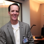
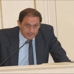
In an exclusive interview, Mauricio Castillo, president of the American Roentgen Ray Society (ARRS), spoke about the impact of dissatisfaction and gender discrimination in radiology, the focus of his Wilhelm Conrad Roentgen Honorary Lecture at ECR 2017.