Hybrid
Automatic image fusion
Dirk-André Clevert MD, Associate Professor of Radiology at the Department of Clinical Radiology, University of Munich, Germany, describes a new diagnostic approach in abdominal ultrasound.
The prevalence of liver lesions is around five percent of the whole population. At the time of diagnosis tumour patients show hepatic metastases in 25-50% of cases and the liver is the main region for metastases in oncological diseases. Therefore, to evaluate the prognosis and establish a therapeutic procedure a reliable detection of hepatic metastases is a prerequisite.
Establishing the number and location of lesions is essential in order to evaluate the prospects of resection, or to plan interventional therapy. Prior to this, the focal hepatic lesion must be accurately characterised. To this end, a second opinion in cases of unclear liver lesions and in cancer follow-up examinations after chemotherapy is extremely helpful and will improve prognoses.
Liver examination using the image fusion technique requires hardware components in the form of a magnetic field generator and an abdominal imaging transducer. Having dedicated positioning software to enable detection and tracking of the transducer is also a necessity. The positioning system calculates the exact position of the respective transducer.
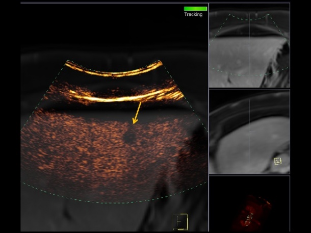
DICOM data of all cross-sectional CT or MRI examinations can be used to upload the images into the ultrasound system for automatic registration. All ultrasound techniques, such as B-mode, duplex-US, power Doppler or contrast-enhanced ultrasound, can be integrated into the image fusion examination. Using the Siemens Acuson S2000 ultrasound system (Siemens Medical Solutions USA, Inc) to use either a conventional magnetic field generator, placed at the patient’s side, or a magnetic field generator integrated into a flat plate placed under the patient, and enables enhanced performance with an innovative automatic registration mode, meaning no manual points or plane registration is necessary. This auto-registration provides extremely fast automatic registration of the data, helping to minimise the exam time and improve the registration quality.
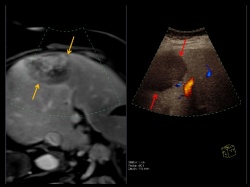
After successful automatic registration and image fusion, the registered CT or MRI images will be simultaneously shown on the ultrasound monitor with the respective ultrasound sectional plane. Additionally, two simultaneous CT or MRI planes could be used to improve visualisation of the lesion.
Image fusion combines the advantages of comprehensive anatomical information due to 3-D CT dataset or MRI dataset and accurate real time dynamic information of contrast-enhanced ultrasound.
Simultaneous observation of the respective CT or MRI images, in the corresponding axial and sagittal planes, improves spatial orientation of the US investigator and helps to accurately describe the exact site of potential lesions or monitor the lesion in the follow-up of the liver. This may be especially relevant prior to therapeutic interventions such as open surgery or radiofrequency ablation.
Image fusion enables direct comparison between CT, MRI and US data and therefore simplifies the follow-up of patients. This is important because enlargement, or the appearance of new liver lesions, is an indirect sign to change the therapeutic strategy.
Regarding vascular diseases or unclear kidney lesions there are a couple of studies showing the advantages of image fusion using CEUS and MS-CT.
26.10.2015