News • Technique comparison
Contrast-enhanced digital mammography vs. breast MRI
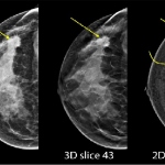
Contrast-enhanced digital mammography is comparable to breast MRI in evaluating residual breast cancer after neoadjuvant endocrine therapy or chemotherapy, according to the results of a study presented by Mayo Clinic researchers at the 2017 San Antonio Breast Cancer Symposium. “Our study aimed to compare contrast-enhanced mammography with breast MRI in evaluating residual breast cancer in…