The ultimate in ultrasound
A joint meeting combining the Euroson and Three Countries congresses creates a veritable European summit on the state-of-the-art in ultrasound


A joint meeting combining the Euroson and Three Countries congresses creates a veritable European summit on the state-of-the-art in ultrasound
‘Cardiology is one of the most innovative medical disciplines. Many modern technologies, such as catheterisations or imaging procedures, were triggered by cardiology,’ declared Professor Dr Gerald Maurer MD.
The future will be aesthetic or, put another way, Art meets Science. With this motto, the 43rd Congress of the German Society for Endoscopy and Imaging Procedures e.V., jointly held in Munich with six other specialist associations, demonstrated that aesthetic means the brilliance of images generated by the latest generation of X-ray, CT, MRI and ultrasound equipment.

CT scanners now nicely cover morphology. The challenge is moving to CT functional imaging without frying patients
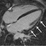
MRI has become the gold standard for many indications in cardiac imaging, apart from imaging the coronary arteries. For function and morphology assessment, MRI is the leading technology. A further advance into as yet unknown territory is myocardial imaging aided by one of the first integrated 3-Tesla PET/MR systems currently used at the Institute of Radiology, Essen University Hospital,…
The new Aquilion ONE ViSION is the widest, fastest, thinnest-slice CT ever built, capable to pushing both anatomical and functional studies to new levels.
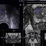
Prostate cancer is the second leading cause of death among men yet remains one of the most frustrating for physicians to find and treat. Merging the strengths of imaging modalities helps, but does not solve all the problems, says John Brosky
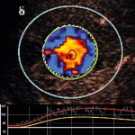
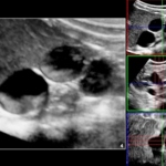
Adding micro-bubbles to the blood lights up the body for ultrasound scanners. Non-invasive, non-toxic and low-cost, these examinations present a disruptive, readily accessible technology for diagnosing disease. John Brosky reports
Dr Peter Choyke, Chief of the Molecular Imaging Programme at the National Cancer Institute in Bethesda, USA, believes that new tracers will have an evolving role to play and represent an exciting development in the imaging of cancers.

Enormous technical developments over recent decades, along with greater understanding of tumour biology, have made imaging, pathology and laboratory medicine indispensable tools in personalised cancer treatments.
Critical ultrasound, as a tool for immediate therapeutic decisions, and emergency POC ultrasound – an extension of the clinical examination at the bedside or on the accident scene – have shown clear benefits along with lung ultrasound.
This may sound like science fiction, but computed tomography with reduced radiation exposure and the highest soft tissue contrast is likely to be a reality -- very soon. Named phase-contrast imaging, the method is an invention of Professor Franz Pfeiffer, Chair of Biomedical Physics at Munich Technical University, Germany. We asked him to explain the implications this development has for…
For the paediatric radiologist an ultrasound system is as essential as a wrench for the mechanic, for three reasons, says Professor Michel Claudon, Head of the Department of Radiology at the Children's Hospital of Brabois, University of Nancy, France: ‘High image quality due to the low weight of children, which allows medium frequency, the absence of radiation and the possibility of performing…
World of Ultrasound met Prof. Ioan Sporea, Prof. Gebhard Mathis and Prof. Byung Ihn Choi to ask them three questions.
Contrast agents have opened up entirely new possibilities are taking shape for ultrasound, above all in oncology. Following the publication of guidelines on the clinical use of contrast-enhanced ultrasound (CEUS) by the European Federation of Societies for Ultrasound in Medicine and Biology (EFSUMB) in 2004 and 2008, at this year’s World Ultrasound Congress, WFUMB and EFSUMB will present joint…
Although many new features in US-guided interventions are being marketed, which are really necessary, which just nice-to-have? It’s a question to be faced by experts during the refresher course ‘Interventional ultrasound’ at WFUMB 2011. One of the most established ultrasound techniques in minimally invasive procedures is contrast enhanced ultrasound (CEUS) – a tool that is safe, gentle…
Around 700 international experts met in Vienna to discuss the latest advancements in ultrasound, such as a new technique called real time imaging, and of approximately 200 scientific papers. That was back in 1969 when for the first time physicians and scientists from around the world came together in Austria’s capital to share their knowledge of the use of ultrasound waves in medicine. The…

Contrast induced nephropathy (CIN) is widely recognised as a potentially serious complication of contrast media use -- a risk that increases with a patient’s age and decreased renal function. Mark Nicholls reports
Medical imaging has recently advanced so rapidly that it should halt. Applying more power to computed tomography (CT) and magnetic resonance imaging (MRI) scanners is becoming too dangerous for patients and healthcare workers. Magnets for the next-generation MRIs are so powerful that they must be moved to a separate building on hospital campuses, while CT radiation levels have risen to alarming…
Michael Maher, Professor of Radiology at the University Collage, Cork, Ireland, produced an answer to during a GE Healthcare Lunch Symposium at the European Congress of Radiology this March. It is 1.2 millisievert – at least for abdominal CT scans of Crohn’s disease patients.
Bayer HealthCare announced that the company will present preclinical data that introduce investigational compounds for both therapy and imaging in its early oncology pipeline with unique and novel mechanisms at the American Association for Cancer Research (AACR) 102nd Annual Meeting, April 2-6, 2011, in Orlando, FL.
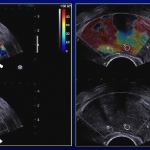
Prostate cancer is the 2nd leading cause of cancer death in men. It is also the most common diagnosed malignancy in men with near 190.000 new cases in the USA in 2008. Despite the larger use of biological tests (such as prostate specific antigen (PSA)) and imaging modalities (trans-rectal ultrasonography (TRUS) and Magnetic Resonance Imaging (MRI)), there is a slight increase in the annual death…
A multi-centre French study is demonstrating that a four-minute ultrasound scan using a contrast agent can be performed after the first month of treatment and provide quantitative proof of whether a tumour is responding to the therapy.
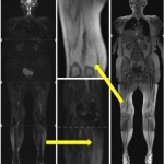
Muscular diseases belong to a heterogeneous group with various causes like neurogenic, metabolic, dystrophic, or inflammatory mechanisms as well as channelopathies leading to disorders of the muscle cell membrane potential. In most progressive disease cases the result is a focal or general muscle weakness that, unfortunately, is a very unspecific symptom. Standard neuromuscular literature…
The 90-minute refresher course ‘Contrast Agent Issues 2010: What the Experts Really Do for Allergies, Contrast-induced Nephropathy, Nephrogenic Systemic Fibrosis, and Extravasation’, to be held during this year’s Radiological Society of North America meeting, will focus on the use of iodinated and gadolinium-based contrast media and the issues, advantages and considerations for patient…