Cardiology drives innovation
A leading Austrian professor commends scanner advances
‘Cardiology is one of the most innovative medical disciplines. Many modern technologies, such as catheterisations or imaging procedures, were triggered by cardiology,’ declared Professor Dr Gerald Maurer MD.
In our EH interview, held during the annual meeting of the Austrian Cardiology Society in June, Prof. Maurer, Head of the Department of Cardiology at Allgemeines Krankenhaus Wien (Vienna’s General Hospital) and Director of the University Clinic Internal Medicine II at Medical University Vienna, outlined the most recent technological developments in cardiology.
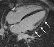
‘MRI images of heart structures are becoming increasingly precise and with increasing resolution,’ he said. This includes the late enhancement, contrast MRI technique, where the contrast agent Gadolinium-DTPA provides detailed information on the metabolism and status of heart muscle cells, supporting tissue differentiation and allows a more precise diagnosis of the cell vitality. The cardiologist can see whether a sub-endocardial infarction occurred where the necrosis, due to a lack of perfusion, is limited to the innermost layer of the heart muscle while the outer layer of the heart muscle is not involved. With myocarditis late enhancement has become the most important diagnostic indicator.
Today in cardiac ultrasound, so-called strain imaging provides new functional information. In 2-D imaging the speckle tracking technology tracks several points in the heart muscle throughout the entire heart cycle and can thus look at the deformation (strain) of the myocardium.
This is particularly relevant in patients with heart failure (HF) with preserved ejection fraction (HFPEF). For a long time cardiac insufficiency was thought to be solely associated with the lack of the heart’s contraction ability. However, around 50% of cardiac failure patients show normal ejection fractions. While some of these patients show a diastolic dysfunction, systolic abnormalities can also contribute. In those patients the myocardial longitudinal shortening is inadequate – a dysfunction that can be detected in strain imaging. ‘In an aging population HFPEF is a growing public health problem,’ Prof. Maurer points out.
He believes that cardiac computed tomography (cardiac CT) will also play an increasingly important role. Cardiac CT visualises the coronary vessels well and helps determine the calcium or Agatston score, an important indicator of a coronary disease. ‘While this method does not replace the catheter, it allows the exclusion of significant coronary heart disease with high probability,’ he explains, adding that a further development is the measurement of cardiac profusion with contrast-enhanced CT.
In the area of cardiac implants, bio-absorbable stents are an important innovation. ‘One of the problems with conventional stents is the fact that in young patients they may be associated with long-term risks. In an 80-year-old patient this is not that much of an issue, but there are many patients between 20 and 40 years of age who suffer CHD and need an implant.’
While conventional stents remain intact for several decades, bio-absorbable stents, which are made from polylactide, dissolve within two to three years – with water and carbon dioxide absorbed by the body. In most cases the endothelium fully recovers and the patient receives a conservative therapy with cholesterol and BP medication and anticoagulants. Large long-term studies are currently being conducted but final results are not yet available.
Profile:
Professor Gerald Maurer MD, who has headed the cardiology department at AKH (General Hospital) in Vienna since 1993, is also Director of the University Clinic Internal Medicine II at Medical University Vienna. He gained his medical degree and doctorate at Vienna’s medical school and completed his specialist physician training in the USA (American Board of Internal Medicine; Subspecialty Board, Cardiovascular Disease). He became a professor at the University of California (UCLA), Head of Non-invasive Cardiology and Interim Head of Cardiology at Cedars-Sinai Medical Centre in Los Angeles before returning to work in Austria. Prof. Maurer is Member of the Board of the Austrian Cardiology Society (ÖKG) and currently Editor-in-Chief of the European Heart Journal – Cardiovascular Imaging.
01.09.2013


