Article • Education
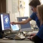
Cardiologists gain MRI training
Seeing a substantially increasing importance of the cardiac MRI procedure, cardiologists have developed a specialist cardiac MRI training programme for their colleagues.


Seeing a substantially increasing importance of the cardiac MRI procedure, cardiologists have developed a specialist cardiac MRI training programme for their colleagues.

At ECR 2015 Esaote, a world leading manufacturer of medical diagnostic systems, introduced Evolution’15 (EVO’15) as the latest upgrade in its dedicated MRI Evolution program. EVO’15 combines software updates and new hardware features to provide superb image quality and increases productivity by almost 50%.
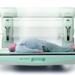
MRI is increasingly relevant to cancer management, especially to detect breast carcinoma. Professor Christiane K Kuhl from the department of diagnostic and interventional radiology at the University of Aachen, Germany, strongly advocated in favour of MRI in breast cancer screening during a dedicated Satellite Symposium organised by Bracco at ECR 2015. Report: Mélisande Rouger
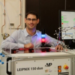
Clinical routine would be inconceivable without MR Imaging. Without exposure to radiation, doctors can make a patient’s organs and tissue structures clearly visible. However, pathological changes in the early stages, degenerated cells or small areas of inflammation, have so far remained almost invisible on these images. In 2014, for the first time, a team of cell biologists, chemists and…

In breast cancer care each patient receives personalised, highly effective diagnosis and treatment procedures. In breast diagnostics this mainly revolves around new MRI scanning procedures that allow the quantification of biological and physiological processes on a cellular and molecular level.
ECR 2015 evoked Shakespeare´s repertoire as delegates were asked to ponder the ethical implications of incidental findings in large population imaging studies in Vienna. Report: Mélisande Rouger

Spring can be felt, though many visitors of ECR 2015 probably only have time to enjoy the floral decorations in the conference center. The program of the conference is packed full of topics that promise exciting days and many discussions.
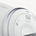
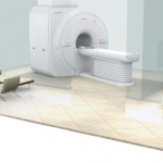
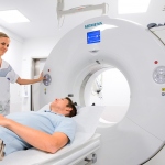
Siemens Healthcare’s new 1.5-tesla scanner MAGNETOM Amira enables clinical excellence with comparatively low costs per scan. MAGNETOM Amira offers the same technologies that are available on the Siemens flagship MRI systems. The new MRI system stands out on account of its lower operating costs.
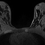
A magnetic resonance spectroscopy (MRS) technique that monitors biochemical changes in tissue could improve the management of women at risk of breast cancer.
At last year’s ECR Toshiba introduced the Vantage Elan 1.5 Tesla system with a lot of innovative features and new techniques, making it a pleasant and helpful new workhorse for small and large clinics. Since this introduction, the Vantage Elan has seen fantastic success in Europe because of the outstanding clinical and economic benefits it brings with advance technologies. The Elan offers…
When the Medical Radiological Institute (MRI) at the private Bethanien Hospital in Zurich and the local hospital in Ærø, Denmark, needed new fluoroscopy and radiography equipment, they investigated quality, functionality, service quality and cost. Among systems examined was Shimadzu’s Sonialvision G4, which has been completely revised, with innovations in all areas, including dose reduction…
Venture capitalists are betting $100 million that an entrepreneur can develop an inexpensive and portable imaging device that can be used by office-based physicians.
A DVD designed to help people prepare for a Magnetic Resonance Imaging (MRI) scan, including guidance on how to relax, led to more successful scans. The patients receiving the DVD also felt less anxious during the scan says a paper published in the British Journal of Health Psychology.
The global Magnetic Resonance Imaging (MRI) systems market will more than double in value from approximately $4.1 billion in 2013 to over $9.2 billion by 2020, representing a Compound Annual Growth Rate (CAGR) of 11.8%, according to research and consulting firm GlobalData.
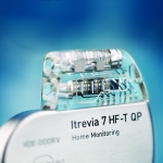
Cardiovascular technology specialist Biotronik has launched a new series of single and dual chamber implantable cardioverter defibrillators (ICDs) and cardiac resynchronisation therapy defibrillators (CRT-Ds). ‘The Iperia/Itrevia/Inventra series gained CE approval in July 2014 and marked its first implantations worldwide in mid-July,’ the multinational biomedical technology firm reports.

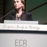
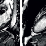
Cardiomyopathy is a disease with many faces, a 'chameleon', according to Professor Jeanette Schulz-Menger. MRI benefits and potential should be communicated better and to a wider clinical audience. Report: Axel Viola

'In paediatric cardiology, echocardiography is the method of choice for preoperative diagnostic purposes,' explains Professor Dr Emanuela Valsangiacomo-Büchel, senior cardiologist and director of cardiovascular imaging at the University Children’s Hospital Zurich, Switzerland. Report: Axel Viola
You are curious to know what this cardiac MRI thing is all about? You want to brush up on your cardiac MRI knowledge? Then we are afraid you have to delve into the technical basics. Sounds boring? It sure isn’t, says Dr Harald Quick.
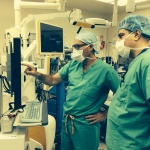
The adage ‘practice makes perfect’ is applicable to every profession – but even more so for pilots and surgeons. Flight simulation technology has been used for decades to hone aviators’ skills, and this technology is now being used by neurosurgeons to plan as well as practise surgical procedures and for real-time virtual assistance in operating theatre. Report: Cynthia E Keen
Andalusia Health Service has selected Accenture and Carestream Health to deploy a picture archiving and communications system (PACS) that will allow clinicians to manage, store and share diagnostic imaging data across more than 1,600 healthcare facilities in Spain. This initiative by the Andalusia Health Service is expected to go live in late 2015, creating one of the largest medical imaging…
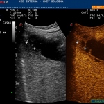
Once considered an add-on examination, CEUS expands diagnostic capabilities and is leading to new insights. Where contrast enhanced ultrasound (CEUS) is restricted to radiologists, or performed only by trained technicians, its utilisation has stabilised, taking its place among more established imaging modalities. Report: John Brosky
Approach could improve treatment of drug-resistant infections. Combining a PET scanner with a new chemical tracer that selectively tags specific types of bacteria, Johns Hopkins researchers - working with mice report - have devised a way to detect and monitor in real time infections with a class of dangerous Gram-negative bacteria.
The significant benefits of cardiac catherisation remain undisputed. However, cross-sectional imaging modalities are serious competitors when it comes to arriving at the right diagnosis.
Imaging has progressed at vertiginous paces since X-rays were invented, not only as a diagnostic tool but also as an invaluable partner in the realm of non-invasive medical intervention.
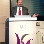
‘There are aspects of the heart’s physiology that we know about, but now we can see them, and this is absolutely different,’ said Patrizio Lancellotti, President of the European Association of Cardiovascular Imaging.