Ultrasound in progress
Ultrasound technology is continuously developing and competing with the sectional imaging procedures – therapy progress can be monitored, facilitating personalised medicine.

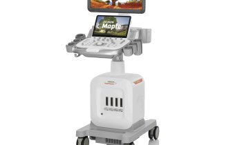
At Heidelberg University Hospital there is excitement about the first use, worldwide, of the latest ultrasound innovation from Siemens. The brand new HELX Evolution not only offers much-improved image quality and far more precise and detailed examination facilities for clinical routine but also, thanks to the special measuring procedure for tissue elasticity, new opportunities to monitor the treatment of cancerous diseases.
The objective in Heidelberg is to use the new scanner to research whether the use of higher-impact procedures, such as CT and MRI scanning, as well as the visualisation of the vessels and ducts with contrast media, can be replaced by much lower-impact ultrasound examinations.
Measuring tissue elasticity with the help of elastography is, in itself not a new procedure, but the HELX Evolution from Siemens also makes it possible selectively to determine tissue elasticity within the ROI (region of interest) after the examination. ‘It is possible to not only measure a certain point while scanning, but also to examine the entire ROI of a size of 6cm x 5cm quantitatively at any time.
‘With current standards, the scanners only deliver a colour-coded map without absolute values, but now it’s possible to extract the absolute value in metres per second at any individual point within the colour-coded map after the examination,’ explains Dr Erick Amarteifio, a senior physician in the Diagnostic and Interventional Radiology Department at Heidelberg University Hospital.
The importance of the actual diagnostic benefit of this absolute value will now be researched. It is known that the speed at which the shear waves spread within the tissue allows conclusions regarding tissue elasticity. The faster the waves spread, the harder the tissue. As many tumours have harder tissue due to high cell density, this can be a first pointer towards a tumorous disease.
One possible application area could be to assess therapy response for hepatocellular carcinoma (HCC) after transarterial chemo-embolisation (TACE). ‘At the moment, the determination of perfusion during an MRI allows conclusions as to the vitality of the tumour. With larger subcapsular foci in particular, we believe it is beneficial to evaluate whether the change of tissue elasticity may allow conclusions as to therapy response with the help of elastography,’ he explains.
This would considerably simplify the progress examination for this group of patients. Instead of a complex and expensive MRI examination, in future it may be possible to monitor and assess a focus with the help of a simple, fast and much better tolerated ultrasound examination. ‘In any case,’ Dr Amarteifio confirms, ‘the HELX Evolution improves the opportunities to further utilise elastography.’
Profile:
Dr Erick Amarteifio gained his doctorate at the Georg-August University in Göttingen and has worked in the Radiology Department at Heidelberg University Hospital since 2008.
At the German Cancer Research Centre, in 2010, he carried out a one-year research project on functional musculoskeletal imaging.
04.03.2014


