Bringing innovative technologies together
Prostate cancer is the most common cancerous disease in men. Since magnetic resonance imaging (MRI) arrived the diagnostic capabilities for early detection have improved considerably, along with more selective prostate cancer treatment.

In particular, the capabilities for tissue differentiation and spatial resolution are much better with MRI compared to ultrasound imaging. However, despite these continuously developing imaging capabilities, the final differential diagnosis and clarification for an MRI result is still a biopsy. The most common type is the ultrasound-guided core biopsy, which systematically samples tissue in 12 locations where prostate cancer most commonly develops. So, why not combine the two technologies, MRI and ultrasound, to reduce the number of biopsies required and achieve higher accuracy with a new procedure?
The Urostation from KOELIS and Samsung Ultrasound
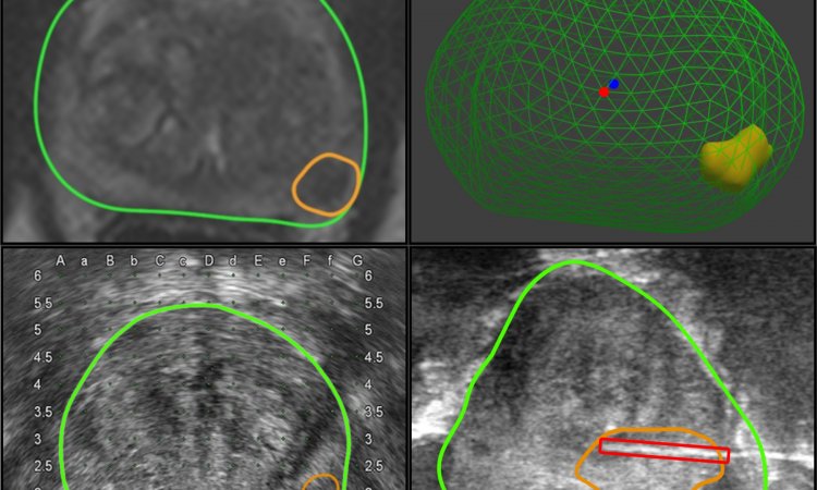
In partnership with the French company KOELIS, the Korean medical devices manufacturer Samsung has developed a urological workstation that combines the advantages of MRI imaging with the practical advantages of ultrasound-guided biopsy. In the Urostation, 3-D MRI images of the prostate are superimposed with live images from the Samsung ultrasound scanner taken with a transrectal 3-D ultrasound transducer.
At the Urology Clinic in University Hospital Dusseldorf, Professor Peter Albers and team are already using a combination of Samsung Ultrasound and Urostation. The clinic is a Prostate Cancer Treatment Centre certified by the German Cancer Society. More than 2,000 in-patients and 7,000 out-patients are examined and treated there every year.
Most biopsies are carried out with the Samsung-Urostation combination. ‘With this procedure we can sample tissue very specifically in locations that look conspicuous on the MRI images,’ Professor Albers explains. ‘The MRI shows up small tissue changes in the prostate at an early stage, which we would not be able to see with ultrasound alone. To take very specific tissue samples, in some individual cases we carry out MRI-guided biopsies.’
However, tissue samples are not only taken from conspicuous locations but systematically from 12 typical locations, although the doctors aim at particularly conspicuous areas. ‘The hypothesis that it suffices to only limit ourselves to particularly conspicuous MRI results has not been scientifically confirmed. Two large studies are currently being carried out to clarify this issue,’ he adds. A welcome side effect: Whilst MRI-guided biopsies are not usually reimbursed by health insurers, this service can now be offered to patients in the university hospital as part of these studies.
More comfort for patients and improved hospital processes
Professor Albers sees the advantages of the new procedure for patients in the method itself. ‘If a patient undergoes an MRI-guided biopsy he has to lie in the MRI scanner on his front for an hour with his arms over his head while we carry out a transrectal biopsy. With the capabilities of the new image fusion, we initially take the diagnostic MRI images and the patient can then lie on his back in the lithotomy position for the biopsy, with the whole procedure over in around ten minutes.’ This not only makes things a lot more comfortable for the patient but also eases strain on doctors.
Hospital processes have also improved through the Ultrasound-Urostation combination. The Urology Clinic shares the MRI scanner with other clinics and the machine is only available for all examinations and biopsies one day a week. ‘The capacity for MRI examinations represents a bottleneck, so we are happy to move the time-consuming biopsies from the MRI scanner to the Samsung Ultrasound-Urostation combination. This gives us more time for diagnostic MRI examinations,’ the professor explains.
Further advantages of the Samsung Ultrasound-Urostation are that examinations and the localisations of biopsies are archived and each localisation can be assigned the corresponding histological result. Therefore, in the case of follow-on examinations, or control biopsies, the doctor can revert to the archived, previous results, inclusive of planning and treatment images as well as relevant diagnostic data.
Choosing the right partner
The combination of Samsung ultrasound scanners with the KOELIS Urostation has proved to be very successful. KOELIS chose Samsung as a partner due to the firm’s high level of innovative technologies, specific design, user friendliness and high quality. The Samsung ultrasound systems SonoAce X8, Accuvix V10, SonoAce R7 and UGEO H60 are already compatible with the Urostation. Further systems, such as the Samsung Accuvix A30 and future models will also be compatible.
Profile:
Peter Albers gained his medical degree at Mainz University (1988) and his urology residency at the Universities of Mainz and Bonn. In 1993-’94 he was research fellow at the Urology Department at Indiana University, USA. Earlier roles also include the chairmanship of the Urology Department at Klinikum Kassel (2003-2008), and Vice Chairman of the Urology Department at the University of Bonn (1998-2003). Since 2008, he has been Professor and Chairman of the Urology Department at the University of Düsseldorf.
18.11.2013


