News • SARS-CoV-2 in the media
Does 'beautifying' the Coronavirus make us underestimate its danger?
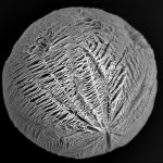
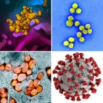
Colourful, 3D rendered scientific images are fascinating - but can they deceive viewers? New research from Spain suggests this might be the case. According to the study by the Instituto de Radio Televisión Española and the Universitat Autònoma de Barcelona conducted during the Covid-19 lockdown, black and white images of SARS-CoV-2 make the virus seem more infectious. The results, published on…