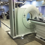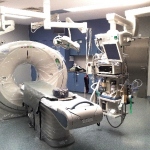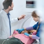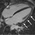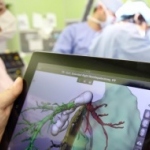
Teleradiology and education
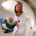
Given the ever more complex radiological examinations, the need to provide care in sparsely populated regions, or new labour law provisions such as the EU working time directive, radiologists are under increased pressure to find solutions to provide imaging services during off-hours.