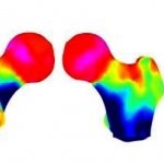
DMS: Breakthrough technology in bone densitometry
DMS (Diagnostic Medical Systems) is thrilled to unveil, for the occasion of the ECR 2013, the newest breakthrough in bone health management: 3D-DXA.


DMS (Diagnostic Medical Systems) is thrilled to unveil, for the occasion of the ECR 2013, the newest breakthrough in bone health management: 3D-DXA.
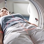
A UK hospital is assessing trauma patients by taking them directly for CT scans rather than to the A&E department. Piloted at King’s College Hospital, this new approach to assessing patients with life-threatening injuries aims to speed up diagnosis by conducting CT simultaneously with patient resuscitation and stabilisation.
CT scanners now nicely cover morphology. The challenge is moving to CT functional imaging without frying patients
For the 19th consecutive year the annual meeting of the European Society of Radiology (ESR), the European Congress of Radiology (ECR), takes place in Vienna.

Mark Nicholls discovers how a CT scan at a British hospital played a critical role in identifying the long-lost remains of a 15th Century English king
Professor Ulrich Linsenmaier, a leading expert in emergency radiology, has highlighted the need for clinicians to read image data rapidly in an emergency department if they are to help improve clinical outcomes for polytrauma patients.
Forty years ago an article was published that would change medical practice. In the British Journal of Radiology, English electrical engineer Godfrey N Hounsfield described how he had made a patient’s brain visible non-invasively by evaluating a large number of X-ray images of the skull taken from different directions.
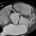
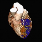
Study described in The New England Journal of Medicine is the first to show cause-and-effect relationship between a gene variant and calcium deposits on the aortic valve.

With a mission to help people avoid unnecessary radiation, and the continuing launch of related products, the Swedish company is now the world’s only provider of comprehensive solutions to measure, monitor and manage X-ray radiation dose, reports Brenda Marsh
A pioneer in lowering patient exposure to radiation, Philips Healthcare opened a new path to further the clinical potentials for CT imaging introducing Iterative Model Reconstruction (IMR) at RSNA 2012.
The new Aquilion ONE ViSION is the widest, fastest, thinnest-slice CT ever built, capable to pushing both anatomical and functional studies to new levels.
– The demand for multi-slice computed tomography (CT) systems will drive growth in the European CT market.
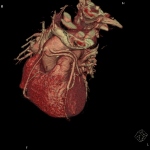
Cardiology is playing an increasingly important role in today’s healthcare environment and, as a direct result, cardiologists are facing new challenges almost every day. Addressing the need of improving clinician confidence and diagnostic accuracy, Toshiba Medical Systems Europe presented two symposia on the first day of the European Congress of Cardiology, to be held in Munich, Germany, 25-28…
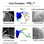
John Brosky reports on a ground-breaking trial and how CT-FFR may change the practice of invasive cardiology and cardiac surgery.
Cardiovascular diseases, the most common cause of death in the West, includes diseases for which early detection is an important objective in cardiac imaging – particularly for coronary artery stenosis. Diagnosis is often made in the cardiac catheter laboratory. Now, however, CT scanning advances provide a lower impact alternative to that invasive exam. PD Dr Thomas Schlosser, Consultant at the…
Incorporating coronary CT angiography (CCTA) into the initial evaluation of low-risk patients coming to hospital emergency departments (EDs) with chest pain appears to reduce the time patients spend in the hospital without incurring additional costs or exposing patients to significant risks. The report of a study conducted at nine U.S. hospitals appears in the New England Journal of Medicine.

When asked about his vision of imaging in the year 2020, Professor Bernd Hamm MD, director of the three radiology clinics at the Charité University Hospital in Berlin, qualified his focus: ‘Technology is always only a vehicle. When we talk about road traffic, we don’t talk about the design of cars but about structural issues’
For his Wilhelm Conrad Röntgen Honorary Lecture at ECR 2011, Professor Richard Baron MD, from the Radiology Department at the University of Chicago, USA, focused on Detecting liver tumours: the search for the Holy Grail. Why does he compare this aim with that of the medieval knights?
Low doses are a major issue among radiologists – but dose management for every single patient, every single system and across modalities is a quite different issue

Despite some decline in cigarette consumption during the last decades, chronic obstructive pulmonary disease (COPD) remains a major public health concern. COPD is among the top five leading causes of chronic morbidity and mortality in the US and in Europe. Nevertheless, COPD is substantially underdiagnosed.
Suspicion of venous thromboembolism (VTE) in a pregnant patient will quickly bring a radiologist to a choice, where the next step holds potentially significant consequences for both the mother and unborn child.
Radiation dose reduction in CT angiography can be achieved by reducing the kV settings, reducing the tube voltage, the tube current and by using iterative reconstruction algorithms.
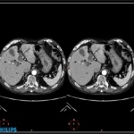
Despite the increasing capabilities of CT to detect or identify disease, fungal infections continue to elude diagnosis by imaging. Paradoxically, a CT examination has been demonstrated to be a powerful tool, helping to identify a probability of infection among immuno-compromised patients early enough to effectively treat the condition.
Will MRI become routine modality? Today, thoracic MRI is rarely performed in Europe. But this will change over the next decade, predicts Professor Hans-Ulrich Kauczor, Medical Director of the Radiology Clinic at University Hospital Heidelberg. He expects Germany to be at the forefront of this development because MRI technology, despite the high costs, is already widely used here and because CT…
“With all that has been published in the past year on this subject, we will not lack interesting things to highlight and discuss during this session,” said Martine Remy-Jardin, MD, Head of Cardio-Thoracic Imaging at the University Centre of Lille, who will present an update on CT Imaging for thromboembolic disease on Friday afternoon at ESTI 2012.