DMS: Breakthrough technology in bone densitometry
DMS (Diagnostic Medical Systems) is thrilled to unveil, for the occasion of the ECR 2013, the newest breakthrough in bone health management: 3D-DXA.
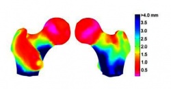
3D-DXA is a brand new technology that allows a 3D model of the femur bone to be constructed from 2D images taken with a DXA bone densitometer during routine femur exams. The resulting 3D model provides key information about the bone status, including bone geometry, cortical bone thickness and bone mineral density.
3D-DXA, a major technological breakthrough in bone densitometry
3D-DXA is the result of a collaboration between DMS, a French company specialized in the development and manufacturing of DXA bone densitometers for the past 20 years, and a leading European research laboratory. The 3D modelization of the femur bone gives specialists access to volumetric slices, allowing for measurements of the bone structure as well as providing volumetric bone mineral density (BMD), information that was previously available only with techniques that deliver higher radiation doses to patients than a DXA exam (a CT Scanner for example). Volumetric BMD along with the cortical bone thickness will help specialists more precisely identify the fragile areas of the bone, thus opening the door to the possibility of anticipating fracture risk, and implementing preventative measures to avoid it.
This technology is currently being clinically evaluated on the femur examination of 120 patients, where the volumetric BMD values and cortical thickness calculated and observed with 3D-DXA are being compared to those calculated and observed with a CT scanner. 3D-DXA constitutes one of the biggest technological advances in the last 10 years in the domain of DXA solutions. Chief Executive Officer Jean-Paul Ansel said, “This work opens the door to a new era, not only in the diagnosis of osteoporosis, but more generally to the diagnostic approach to bone pathologies and bone health evaluation. DMS would like to extend this research to other skeletal sites, with particularly interesting perspectives in fracture risk anticipation and the evaluation of treatments. 3D-DXA allows bone exams to be taken at less cost and with less radiation dose than other current solutions. We have decided to orient our R&D teams at DMS toward 3D reconstruction, which will bring new cutting edge technologies to specialists interested in bone health.”
For further information about 3D-DXA, please watch the film on our website: www.dms.com
09.03.2013