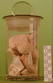
Keep the stones rolling: CT improves diagnosis of kidney stones
It is an every-day occurrence in any emergency department: patients presenting with severe flank pain. In roughly 50 percent of these cases, the pain is caused by a stone. 15 percent of all men and six percent of all women suffer from stones in kidney, ureter, bladder or urethra at least once in their lifetime

At the European Congress of Radiology (ECR 2013), which is currently taking place in Vienna, one session was dedicated to the diagnosis and treatment of such concrements.
Ultrasound is losing ground in the diagnosis of urinary stones. “Never trust a normal ultrasonography,” warns Professor Anders Magnusson of the Department of Radiology at Uppsala University Hospital in Sweden. And Dr. Sami Moussa, Consultant Uroradiologist at the Scottish Lithotriptor Centre (SLC) in Edinburgh, Scotland, adds: “In the past ten years computed tomography is increasingly being used to diagnose stones, but also for follow-up and to guide interventions.”
Ultrasound has two major advantages: it is easily available in every hospital and it does not expose the patient to radiation. But it has also two major drawbacks: poor sensitivity – only 13 percent of stones that are 3 mm or smaller are detected – and strong operator dependence which means that the scanning results depend to a large extent on the experience and the dexterity of the scanning physician. Computed tomography (CT) on the other hand “allows immediate diagnosis with high sensitivity and specificity,” underlines Professor Gertraud Heinz-Peer of the University Clinic for Radiodiagnostics at the Medical University Vienna. Special CT systems can help identify the chemical composition of the stone, be it calcium oxalate, urate or struvite to name the most frequent substances. Dual energy CT uses two different energy spectra for image acquisition while dual source CT simultaneously applies two x-rays positioned in a 90° angle.
Approximately 85 percent of all stones that are smaller than five mm pass out of the body easily. As soon as the stone gets stuck in the ureter however, the flank muscles painfully contract in waves – hallmark symptoms of a renal colic. If the blockage in the ureter leads to an obstructive uropathy, pyelonephritis and uremia can ensue. “The treatment of this pathology is defined by many factors such as the size and the location of the concrements or the patient’s overall condition,” Moussa says. At the Scottish Lithotriptor Centre the most frequently applied therapies are extracorporeal shock wave lithotripsy (ESWL – 82 percent), ureterorenoscopy (URS – 12 percent) and percutaneous nephrolithotomy (PCNL – six percent).
Stones that are smaller than 20 mm are usually crushed with ESWL or URS. If the stones are between 20 and 25 mm ESWL with a so-called double J stent is the therapy of choice. Even larger stones are removed with PCNL. According to Moussa today the percutaneous endoscopic removal of kidney stones is frequently done with the patient in supine position.If the stone obstructs urine flow immediate drainage of the bladder via catheterization is indicated. “If a patient presents with flank pain and signs of an infection an obstruction of the ureter is very likely and a nephrostomy has to be initiated at once,” Magnusson emphasizes, adding that this can be a life-saving procedure. “A ‘stone attack’ is very painful,” the Swedish radiologist reminds us, “but it does not have to be lethal.”
10.03.2013


