Article • Elastography
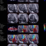
3-D Endocavity probe knocks for targeted prostate procedures
The prostate remains the only organ where random biopsies are performed to find cancer, notes Jean-Michel Correas MD PhD, from the Necker University Hospital in Paris. If we proposed this approach to a woman to search for breast cancer, it would be outrageous, he said.