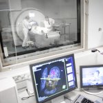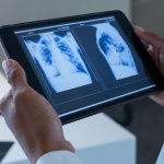
News • In Southern Denmark
Philips empowers image access for over 5,000 clinicians
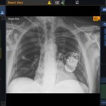
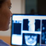
oyal Philips announced the completion of a regional informatics project that consolidates radiology and nuclear medicine imaging data. The Region of Southern Denmark now has a single system for storing, retrieving, and viewing clinical images across all the locations and specialties in its extensive healthcare system.