Article • Orthopedic surgery
The knee – correlations of MRI and arthroscopy
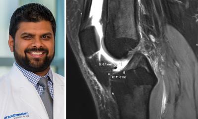
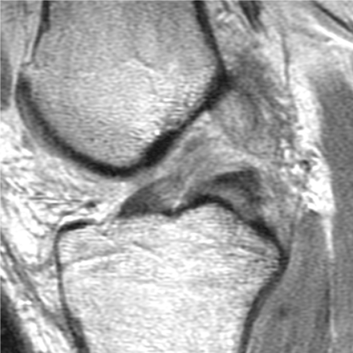
Magnetic resonance imaging (MRI) exams of the knee are essential to orthopedic surgeons for diagnosing the cause of symptoms in patients with knee pain and planning arthroscopic treatment. Yet the surgeons who treat patients based on knee MRIs and the radiologists who interpret those knee MRIs often work in their own silos of specialization, rarely communicating and sharing information, according to William Palmer, M.D., Director of Musculoskeletal Radiology & Intervention at Massachusetts General Hospital, a teaching hospital of Harvard Medical School in Boston.

This lack of communication puts radiologists at a learning disadvantage. Although radiologists review many MRI studies of the knee and report findings, including meniscal tears and cartilage defects, they may not receive feedback on the accuracy of their interpretations. A meniscus that a radiologist reports as torn may be diagnosed as normal at arthroscopy, or a meniscus that a radiologist reports as normal may be diagnosed as torn at arthroscopy. Without feedback from surgeons or access to arthroscopy notes, radiologists work in a vacuum. They may fail to learn about their mistakes and, therefore, are more likely to repeat their diagnostic errors.
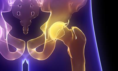
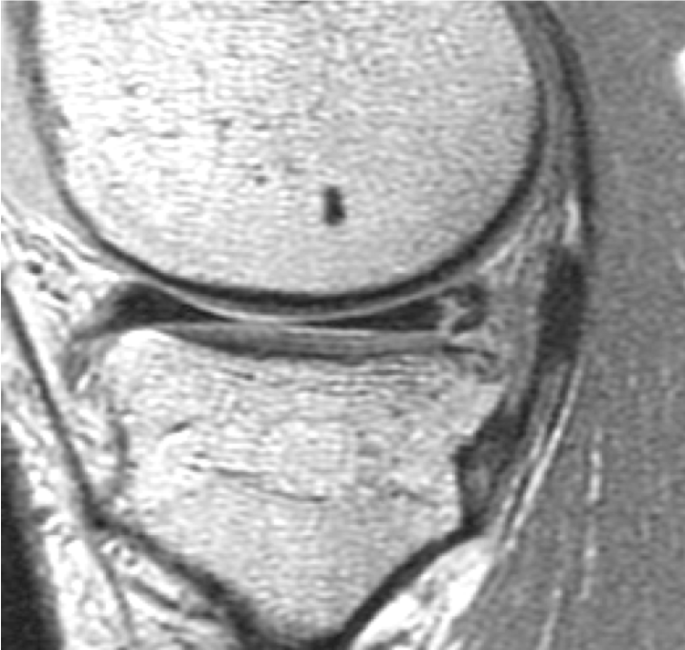
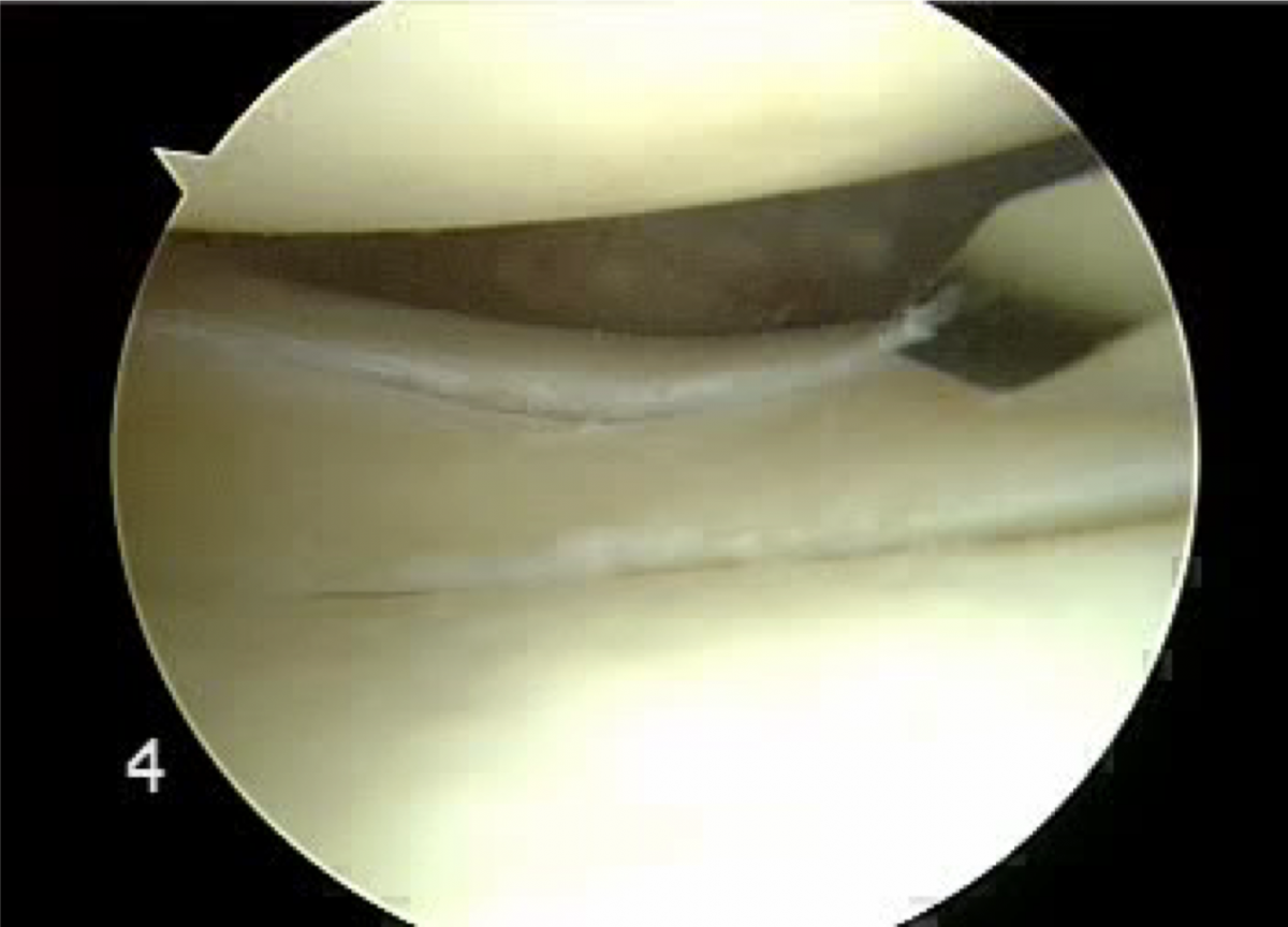
Because arthroscopy is the gold standard in the diagnosis of internal derangements of the knee, Dr. Palmer was asked to give a talk entitled “Knee: MRI - Arthroscopy Correlation” at the 18th MRI Symposium in Garmisch. Dr. Palmer plans to use a biomechanical approach to MRI interpretation in the setting of knee trauma, focusing on sports-related meniscal tears and anterior cruciate ligament injuries. He will compare the MRI and arthroscopic appearances of knee structures and internal derangements.
Dr. Palmer states that “This comparison can be a powerful educational tool. By correlating MRI findings with arthroscopy of the knee, I hope to teach radiologists about patterns of injury and the diagnosis of subtle but important structural abnormalities.”

Profile:
William Palmer, M.D. is Director of Musculoskeletal Radiology & Intervention at Massachusetts General Hospital (MGH). He received his M.D. from Yale University in 1984, and obtained board certification in Internal Medicine (1987, Hospital of the University of Pennsylvania) as well as Radiology (1991, MGH). He completed fellowship training in MRI at Massachusetts General Hospital (MGH) and joined the MGH radiology staff in 1991. He became its Director of MRI in 1995. Clinical expertise includes sports imaging, arthritis and spine intervention. He oversees 9 MSK radiologists and 5-7 fellows.
Sesssions:
Freitag, 18.01.2019, 08:20–08:50 Uhr
MRT des OSG
William Palmer, M.D. (Boston)
Session: Muskuloskelettale Bildgebung
Freitag, 18.01.2019, 14:20-14:40 Uhr
Das Knie: Korrelation von MRT und Arthroskopie
William Palmer, M.D. (Boston)
Session: Film-reading und special focus sessions: MSK & Abdomen
16.01.2019