News • New X-ray technique
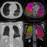
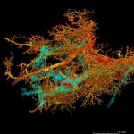
HiP-CT reveals link between long Covid and pulmonary fibrosis
A new X-ray technology has been used to identify a link between the damage that severe Covid-19 can inflict on lungs and pulmonary fibrosis, a disease that causes severe scarring of lung tissue.