Phase-contrast imaging will revolutionise X-rays
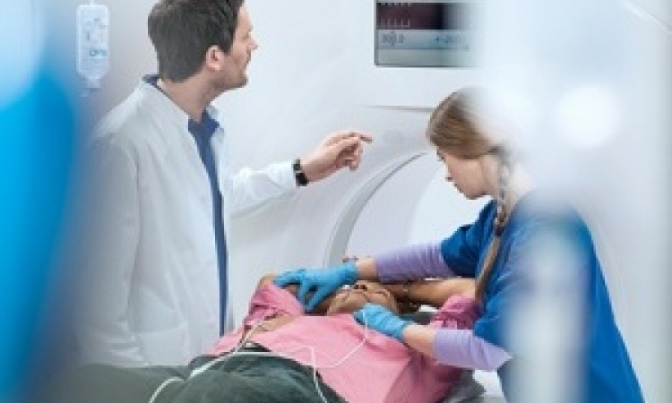
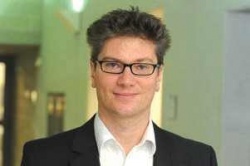
This may sound like science fiction, but computed tomography with reduced radiation exposure and the highest soft tissue contrast is likely to be a reality -- very soon. Named phase-contrast imaging, the method is an invention of Professor Franz Pfeiffer, Chair of Biomedical Physics at Munich Technical University, Germany. We asked him to explain the implications this development has for radiology.
The X-ray physics behind phase-contrast imaging has long been known: X-rays are nothing more than high-energy light and can thus be described as quanta and waves. Waves not only ‘get stuck’ in matter, they also interfere. ‘Just like light that is refracted by a lens, X-ray waves can be refracted by structures,’ Professor Pfeiffer explains. ‘As far as clinical applications are concerned this means that refraction in tissues differs depending on tissue density. Since the angles of refraction are so minute they were invisible in conventional X-ray imaging, but recently we learned to make these tiny refractions visible.’
This visualisation is made possible by small grate-like structures placed one behind the other, which allow identification of the part of the beam that is being refracted. Since physicists speak of refraction in a phase, the term phase-contrast imaging was coined.
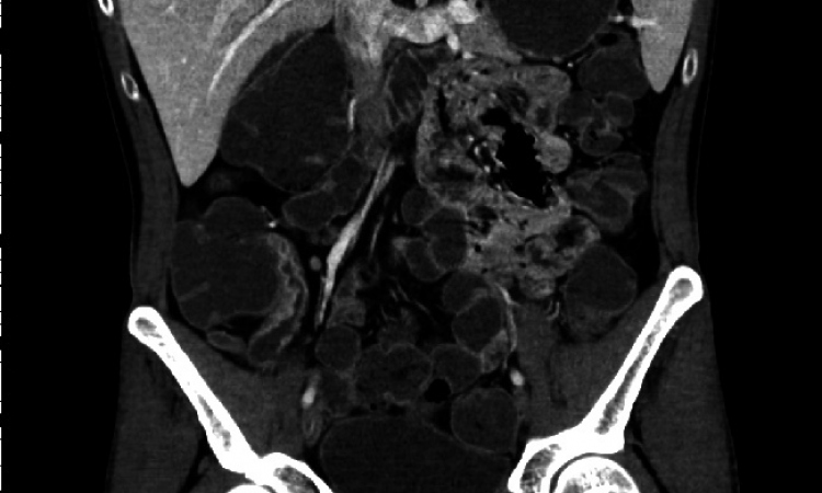
This imaging method is based on the modification rather than absorption of X-rays in tissue in order to create contrast -- thus the quality of the contrast is not necessarily linked to a dose that ‘gets stuck’ in the body.
This is not Prof. Pfeiffer’s only crucial insight. He and his team also discovered that phase interaction generates significantly more contrast in soft tissue. ‘Conventional absorption provides only weak signals in the soft tissue since tissue does not absorb the radiation very well. However, in phase-contrast imaging this limitation does not exist although we are not quite sure yet what – from a bio-medical point of view – creates the soft tissues contrast. Nevertheless, the result is amazing.’
To explore the basics and potential of this novel imaging method, Professor Pfeiffer and team are working on tissue samples received from the radiology departments of Munich’s Ludwig Maximilian University and the Technical University, which have closely cooperated with the researchers for about 18 months. ‘Without the help of radiologists who, after all, will be the end users of the new method, we would not get anywhere’, the professor said. ‘They tell us what they need and where they see problems – things of which we physicists are not necessarily aware. On the other hand, our clinical colleagues have recognised the enormous potential of phase-contrast imaging, for example in early detection of tumours, and thus their interest in having bio-medical basic research translated as quickly as possible into clinical practice is growing.’
This increasing interest is already visible at an international level. For a long time there were only two research groups – one in Japan, one in Switzerland, headed by Dr Pfeiffer – but now there are 19 teams worldwide dealing with phase-contrast imaging.
However, the practical implementation of the new X-ray method is no easy task. Presently, one of the physicists’ biggest challenges is visualisation of the refraction of the X-ray beams to an extent where they can be turned into reliable signals. ‘That’s because of the gratings’, Prof. Pfeiffer explains, ‘they don’t work very well yet for the high X-ray energies in a modern CT scanner.’
It will thus be a while before the current experimental systems are transformed to commercially available products -- but Professor Pfeiffer is ploughing ahead. He is in the process of setting up a CT scanner for small animals, first to perform in vivo studies. Results from the mouse model will be important to convince potential industry partners to team up with the researchers for the next stage of development -- building a CT system for humans. This can only be realised in cooperation with a large company.
Prof. Pfeiffer has no doubt that the interest is there. ‘It’s a bit like building an entirely new engine for a car. At first no one really dares to do it. But, as pressure is mounting so the willingness to invest in innovative top technology grows.’
Who will go for it? We’ll keep you posted!
14.02.2012