Contrast-enhanced ultrasound - the benefits for children
For the paediatric radiologist an ultrasound system is as essential as a wrench for the mechanic, for three reasons, says Professor Michel Claudon, Head of the Department of Radiology at the Children's Hospital of Brabois, University of Nancy, France: ‘High image quality due to the low weight of children, which allows medium frequency, the absence of radiation and the possibility of performing an examination without sedation or anaesthesia due to real time imaging.’
For European Hospital he outlined how children might benefit from the latest ultrasound advances, including contrast enhanced US.
Are developments in ultrasound, such as 3-D, contrast agents or elastography, part of your daily work with small patients?
Professor Claudon: ‘Elastography is not used that much in children. Firstly, we already have easy access to most of the organs. Secondly, tissue in children is often too soft to be characterised based on palpation. And finally, the most important purpose of elastography in adults, namely the characterisation of stiffness of the tissue to detect cancer, is more or less superfluous in children because tumours in young patients are often quite big and therefore easy to differentiate from the surrounding tissue.
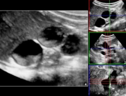
‘However, contrast-enhanced ultrasound is very useful in paediatrics. There are two major indications: The detection and follow-up staging of reflux between bladder and the upper urinary tract, which is quite common in children. Secondly, there are classical indications, such as spleen or liver trauma and the characterisation of focal liver lesions. In those cases contrast agents would be very helpful, but there is a dilemma: Sonovue, the only ultrasound contrast agent available today in Europe, is not approved for children. This means that physicians are forced to use it off-label, with the consent of the parents.’
Are there efforts to find a solution and ‘legalise’ its use for children?
Professor Claudon: ‘Of course; several organisations are working on a solution. At the meeting of the International Society of Paediatric Radiology in London, last May, representatives from the European Society of Paediatric Radiology, the European Society of Uroradiology and the European and World Federations of Ultrasound in Medicine and Biology discussed the possibilities of beginning the approval procedure with the manufacturer of Sonovue (Bracco, Italy).
‘There already are various clinical results regarding CEUS. On behalf of the European Society of Paediatric Radiology, Dr M Riccabona, from Austria, conducted a survey that indicated more than 4000 contrast-enhanced examinations were performed on children during recent years throughout Europe.
‘These mainly concern reflux, but also intravascular indications. No adverse events were reported. This is good news and we think we should proceed with the approval process, although we know it will take time. The first step – putting the topic on the agenda – has been done and we now have to define how to get the European authorities on our side.’
What about volume imaging in ultrasound?
Professor Claudon: ‘Volume scanning has developed from a nice toy to a clinically valuable method. And, with the introduction of purely electronic probes (matrix technology) without mechanical motion it’s becoming even better. The scanning time decreased from a few seconds to only fractions of a second. It is much more efficient and at the same time offers improved image quality.
‘The issue that’s now being debated is how we proceed from there. Do we just add volume acquisition to the 2-D image? This is possible and clinicians would benefit from this new method, for example by calculating the volume of an organ. Or, do we use 3-D as a complementary tool to 2-D examinations? The third alternative could be to acquire volume images and assess them afterwards, as we do with CT scans today.
‘Of course this shift will change our workflow because we’ll no longer see the result of the examination immediately. Volume scanning would mean examining the patient, reviewing the images and then making a diagnosis. This additional time would slow down the workflow but it would offer additional diagnostic options.
‘In view of these developments I’m sure that 3-D will play an important role in ultrasound in general in the future -- particularly in paediatric ultrasound.’
26.10.2011