News • Breast cancer
MR spectroscopy shows precancerous breast changes
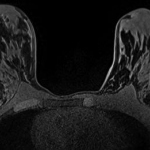
A magnetic resonance spectroscopy (MRS) technique that monitors biochemical changes in tissue could improve the management of women at risk of breast cancer.


A magnetic resonance spectroscopy (MRS) technique that monitors biochemical changes in tissue could improve the management of women at risk of breast cancer.
At the recent North American IHE Connectathon in Cleveland, Sectra (STO: SECT B) successfully completed testing of the IHE profiles MAMMO and DBT (Digital Breast Tomosynthesis). The testing was done using Sectra PACS which implements the Image Display and Image Manager actors of these profiles.
Carestream will show enhancements to its digital breast tomosynthesis (DBT) module including a slabbing tool, improved workflow capabilities and the display of DICOM-compliant 2D synthetic views (which are generated from the 3D dataset) at the 2015 European Conference of Radiology. The latest DBT module is currently available.
Women with metastatic breast cancer know they have a slim chance of long-term survival. The question is whether personalised therapy could extend their lives. Report: Cynthia E Keen
A panel of experts has recommended that existing programmes be phased out and that systematic screening programmes be replaced with systematic screening information that would give women the opportunity to make individual choices. Report: Mark Nicholls
A new study has suggested that mammography screening of healthy women can help to significantly reduce deaths from breast cancer. Much will now depend on new treatments and more systematic management of patients. Report: Mark Nicholls

Expert estimates suggest that women eating more poultry, fish, nuts and legumes and less red meat might have lower risk.

Created by the Dutch industrialist family Hilekes, in 1993 Medicor expanded beyond the Benelux countries to enter the German-speaking world as Medicor Germany GmbH, selling contrast media injectors for CT, MRI and angiography.
Curie-Cancer, the body which leads the Institut Curie's industry partner research activity, and Servier, today announce that they have renewed their partnership with the aim of identifying therapeutic targets for treating ‘triple negative’ breast cancers. The partnership will continue for a further three years.
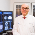
Why is early detection of breast cancer so important? Professor Dr Walter Heindel, Director of the Institute of Clinical Radiology at University Hospital Münster, Germany, offers an unequivocal answer.

Breast cancer hurt her but after a long treatment, she is now 10 years past the day she heard "You have cancer."
It may well be the first computer-aided detection (CAD) software that went to school with radiologists to study breast cancer.
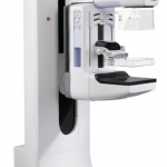
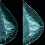
In May, 2013, the U.S. Food and Drug Administration (FDA) finally took the training wheels off tomosynthesis by approving the use of Hologic's new C-View 2D imaging in place of conventional 2D mammograms previously required as part of a breast tomosynthesis screening exam.
RSNA opens a window for one week where companies can showcase the latest technologies ahead of regulatory approval. Fujifilm seized this opportunity to introduce the leading edge in tomosynthesis, the Amulet Innovality that it has launched in Europe, and that once given the green light by the FDA will come to America under the name Aspire Cristalle.
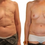
In France, every year 15,000 women undergo complete or partial mastectomy due to breast cancer. Only about a third of them, i.e. around 5,000 patients, use the possibilities reconstructive surgery offers and 70 percent of those women opt for an implant although it is associated with a risk of infection because the body might react negatively to the foreign object.
Toshiba ultrasound equipment is helping provide a better oncological and cosmetic outcome for women recovering from breast cancer treatment and surgery. Daniela Zimmermann discussed intraoperative ultrasound-guided breast surgery with surgeon Dr Monique Petrousjka van den Tol, from the Department of Surgical Oncology, VU University Medical Centre in Amsterdam, the Netherlands

Like any other cancer – breast cancer is a highly individual disease, shaped by many factors such as age, health status or genetics. Due to the complex web of molecular pathological processes and resistance mechanisms it is very difficult to select the most effective therapy for each patient.
Dr Martínez Miravete didn’t set out to change breast imaging in Spain when she first adopted breast tomosynthesis.

Research using an analytical health economics model has suggested the current system of screening within the UK’s National Health Service (NHS) is only moderately likely to be cost effective.
Ultrasound may be used during breast conservation surgery, to locate tumour lesions or to place localising wires; it can also guide a lumpectomy and perform a specimen exam to ensure a lesion has been excised and to evaluate surgical margins

With the help of a commercially available CAD (computer-assisted diagnosis) programme, MRI can provide prognostic data on the development of distant metastases in the further course of breast cancer.
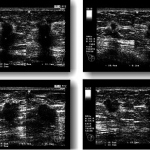
Over the decades of breast imaging numerous studies have shown that radiation free and inexpensive ultrasound can detect some subtle cancers not visible on a mammography exam.

The charity Cancer Research UK reports that the number of breast cancer diagnoses in under 50-year-old women each year in the UK has exceeded 10,000 for the first time.

Self-guided study of more than 6,000 ultrasound exams are at your fingertips thanks to an eight-year voluntary effort by a Dutch radiologist and technical support provided by Hitachi-Aloka.
Israel - Researchers are using breath-test technology to detect volatile organic compounds to tell whether a patient has stomach cancer.