Tomosynthesis proves advantageous in breast imaging
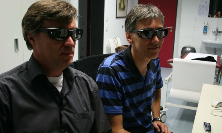
Two years ago Dr Michael Michell and team at King’s College Hospital, London, set out to explore the benefits of tomosynthesis over conventional 2-D mammography. Their study has shown advantages in diagnostic accuracy and indicates that tomosynthesis could help to reduce the number of patient recalls for further examination and thus anxiety among women.

‘We started evaluating tomosynthesis in January 2009 because we became aware of the technology and were fortunate enough to be provided a machine to do some evaluation by Hologic,’ explained consultant radiologist Dr Michael Michell, Clinical Director of the King’s College Hospital’s Breast Screening Programme and National Training Centre. ‘Our pilot study covered 750 patients who had routine screening but had been recalled because something abnormal was found on the mammogram. We wanted to study a population that had quite a large number of abnormal findings.’
The hospital’s breast screening service covers 220,000 women aged 50-7- years in south-east London, carrying out routine mammography examinations on over 50,000 women per annum. The National Training Centre provides specialist training in all aspects of breast radiology for radiologists, radiographers and other professionals involved in the service.
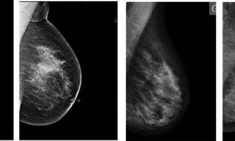
The team’s research has been positive, and has been reported at various meetings in Europe and North America over the past year or so. ‘We found that there seemed to be an advantage in diagnostic accuracy to tomosynthesis compared to 2-D imaging, both in the ability of the radiologist to diagnose cancer and in their ability to diagnose either benign or normal findings,’ Dr Michell said.
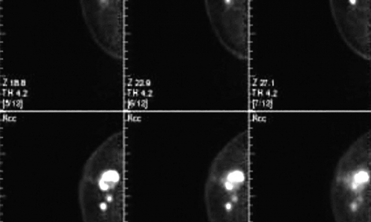
Tomosynthesis is not routinely used at this stage in the UK but is currently being used in his department for the diagnostic workup, along with ultrasound, biopsy and clinical examination. The team is now focusing on how tomosynthesis could be applied in screening practice.
According to Dr Michell, three clear questions need answers: Does tomosynthesis improve the ability of the reader to detect cancer? Does it have the ability to say something is either definitely normal or benign and not cancer? Are there clear cost benefits?
Currently, in the UK and elsewhere in Europe, all screening mammograms are read independently by two separate readers, which is very costly. ‘So, if we could achieve the same sensitivity and ability to detect cancers with one reader, clearly that would be a cost saving.’
However, in terms of accuracy there could be clear benefits, particularly in reducing unnecessary anxiety to women. In the UK, following routine breast screening between 90,000 and 100,000 women are recalled each year for further tests and further workup because results are not clear, but the majority do not have cancer. Figures from 2007-08, for example, show that of the 78,900 women screened in England and then recalled for assessment, about 64,000 were found not to have cancer. In terms of their inevitable anxiety, ‘If we could reduce that number,’ he said, ‘it would be a huge advantage.’
Tomosynthesis might provide better information about the tissues and benign lesions, he pointed out, adding that if the number of recalls of those without cancer could be reduced it would not only be good for the women but also healthcare costs. Once the benefits of tomosynthesis have been established, a closer focus on those costs and the overall cost of the technology could be made, for example, whether it takes longer to read tomosynthesis images or the 2-D images, or whether that cost is offset by having fewer readers and less assessment or diagnostic workup clinics.
‘It’s about balancing the funds you have and ensuring you use them most efficiently -- a challenge for any healthcare programme in the world, particularly in Europe at the moment,’ he added.
Tomosynthesis is a relatively new technology, and there are differences in the appearance of the 3-D images to the 2-D images. Dr Michell has established a training programme to enable radiologists to work safely and ‘use the technique to advantage of patients’. The training package was developed from some of the 750 cases in the London study. ‘We have extracted sets of interesting, difficult and subtle cases, which are useful for training,’ he explained.
Those experiences have now been shared with radiologists in countries that include France, Switzerland, Italy, Holland, Belgium, Germany, Turkey, Israel and Spain.
05.12.2011