From punch to vacuum to RF-guided breast intervention with ultrasound
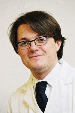
More than 90% of patients who present with suspect or highly suspect breast lesions (BI-RADS categories 4 or 5) now undergo biopsies. Further treatment is only carried out after the precise histological clarification of the tissue sample is obtained. The intervention is routinely carried out via ultrasound guided punch biopsy. Thanks to new techniques the procedure is now becoming even more reliable and can sometimes even completely replace surgery. Professor Thomas Helbich, Director of the Division of Molecular and Gender Imaging, and Vice Chairman of the University Clinic of Radiodiagnostics, Vienna, will report about the different forms of ultrasound-guided biopsy
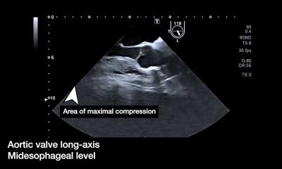
‘Each breast lesion that can be shown with B mode ultrasound can also be biopsied via ultrasound,’ explains Prof. Helbich explains. ‘The image resolution is so excellent that the false negative rate is only 1.5%. The big advantage during the intervention is that ultrasound as real-time imaging also makes the lesions clearly visible during the procedure.’
Next to fine needle aspiration, where only lymphatic fluid is taken (e.g. from cysts) ultrasound-guided punch biopsy is the most commonly used procedure for the extraction of tissue samples. ‘The breast is a very robust organ, with few risks of complications and a low risk of bleeding. We can therefore safely use larger hollow needles than those used for the kidney and liver, for instance. We mostly use 14 G needles with a diameter of 2mm.’ Only in a few exceptions where the lesion is very close to the chest wall is punch biopsy avoided as there is a risk of injuring the thorax with the needle.
Although ultrasound-guided punch biopsy is generally very reliable, the procedure has some disadvantages for some types of lesions, specifically including ‘soft’ lesions. ‘The tissue sample taken may not be reliable enough because it only represents part of the entire problem. This means that, although you may have a very precise diagnosis for the particular sample, the diagnosis may not be the same once the entire tumour is later surgically removed and histologically examined. This so-called underestimation rate is currently around 20%,’ he points out.
A ductal carcinoma in situ may, after surgery, turn out to have been an invasive carcinoma, or an atypical lesion may have to be upgraded to a carcinoma.
For this reason there are efforts to increase the number of ultrasound-guided vacuum biopsies. This procedure not only requires larger needles -- diameter of 3-4mm -- but also means that through the combination of cutting and aspirating technology, larger tissue elements can be extracted from the lesion.
Tissue is sucked into the needle by vacuum and then cut by small knives in the needle. The method works so successfully that there are already trials to see whether benign lesions can be entirely removed by ultrasound-guided vacuum biopsy, sparing a patient from surgical intervention.
A new promising procedure, where the lesion is not cut into small pieces and each individual piece of tissue is individually transported outside, is radio-frequency ablation, in which a system of needles is positioned in front of the lesion through a thin puncture in the breast. A small metal ‘basket’ is then put over the lesion, cuts it from the surrounding tissue, and basket and contents are then retracted from the breast.
‘Here, we are basically at the interventional interface between biopsy and treatment,’ Prof. Helbich point out. ‘Positive results should, however, still be treated with caution. Whether or not radio-frequency ablation really delivers what it promises will be seen in the coming years.’
"Update of US-guided breast interventions", Friday August 26, 14:30-15:00, Hall NO
Thomas Helbich
Between 2007 and 2008 Professor Thomas Helbich headed the Breast Imaging Department -- one of the largest centres for breast diagnostics worldwide -- in the University of Toronto.
In 2008, he took up his role as Professor of Molecular Imaging at the University Clinic of Radiodiagnostics in Vienna, along with that of Vice Chairman of the Clinic. Currently, he is also President of the Austrian Society of Senology and the European Society of Breast Imaging.
The professor’s main research area is molecular imaging with the focus on gender imaging.
25.08.2011