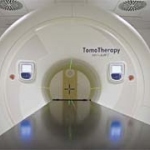
Helical tomotherapy
By Rudolf Schwarz and Andreas Krüll, of the Section of Radiation Oncology Department, Ambulanzzentrum GmbH of the University Medical Center Hamburg-Eppendorf


By Rudolf Schwarz and Andreas Krüll, of the Section of Radiation Oncology Department, Ambulanzzentrum GmbH of the University Medical Center Hamburg-Eppendorf

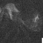
Molecular imaging aims at the in vivo quantitative visualisation of molecules and molecular events that occur at cellular level. The potential towards clinical translation is huge, because the same modalities used in medical imaging are used in molecular imaging investigations.

Imaging in Internal Medicine is among the main topics for 114th Congress of the German Society of Internal Medicine (March. Wiesbaden). Specialists in internal medicine, radiologists, and nuclear medicine have developed a programme that will not only provide an overview of the values of modern imaging procedures but also tackle controversial subjects.

For individualised radiotherapy, high-precision delineation and characterisation of the tumour is critical. If highest radiation doses are delivered in a targeted fashion, the chance of tumour cell kill increases and tumour control probability is enhanced.

Breast cancer morbidity has been the leading oncology disease (21.8%) in Russia since 1996 - and since 1981 in St. Petersburg. In Moscow, the morbidity has increased 52.4% in last 14 years.
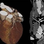
Competitive or complementary? By Florian Schwarz BS, Balazs Ruzsics MD PhD and U. Joseph Schoepf MD, of the Radiology and Medicine Departments, at the Medical University of South Carolina, Charleston, USA.
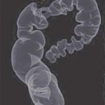
When the International Agency for Research in Cancer (IARC) 2007 statistics report, showed that 429,000 new cases were reported in Europe in 2006, Director Peter Boyle recommended that colorectal cancer screening programmes be implemented throughout Europe.
By Professor Robert D Speller, Head of the Radiation Physics Group, University College London, and Dr Alessandro Olivo, of the Medical Physics & Bioengineering Dept. University College London

The healthcare system is in a phase of transition - from planned economy to free market economy. Competition is becoming a challenge. Only entrepreneurs and enterprises that develop creative strategies will stay on top - or make it to the top.
Agfa Healthcare's new IMPAX solution suites offer PACS and RIS to cover hospital data handling and cardiovascular, cardiology, orthopaedics, mammography and radiology data.
PET/CT imaging exhibits significantly higher sensitivity, specificity and accuracy than conventional imaging when it comes to detecting malignant tumours in children, according to research published in the Journal of Nuclear Medicine (12/07).

Apart from spending a year at Johns Hopkins University Hospital, Baltimore, USA, the professor has never worked anywhere other than at the Radiology Department at the University Hospital of Cattinara Hospital, Trieste, of which she is Chairman. Daniela Zimmermann asked her about what women can achieve in this field, as well as the professor's own multifarious roles and research activities.

Professor Rémy-Jardin MD PhD heads the Department of Radiology and is Chairman of the Department of Thoracic Imaging at the Calmette Hospital, University Centre of Lille. She is also Professor of Radiology in Lille University's Medical Faculty.
US launches a national initiative By Cynthia E Keen

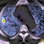
Today, Real-time Tissue Elastography (HI-RTE) provides a method to determine tissue elasticity of certain organs, such as the prostate, in real-time and to perform precise biopsies for reliable tumour diagnosis during standard examinations.

Advances in prostate cancer imaging spark hopes for better therapies. Meike Lerner asked Professor Hartmut Huland, Medical Director of Martini-Klinik in Hamburg, and pioneer of the nerve-preserving prostatectomy method, currently the gold standard in prostate cancer therapy, about his technique and whether the optimism regarding imaging is justified or misplaced

Healthcare is in a dynamic state of change — and so is the healthcare industry, in which there is an increasing trend towards integrating scientific disciplines.
A multi-institutional study recently compared whether positron emission tomography (PET) or computed tomography (CT) performs more efficient in the characterization of solitary lung nodules (SPNs). Previously performed studies were either limited by small sample sizes or carried out more than a decade ago with outdated technology and methods.
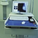
A promising mammography screening technology By Andrew Smith PhD, principal scientist at Hologic, Inc. in Bedford, Mass, is involved in research and development of digital imaging systems.
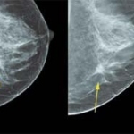
A study recently published in the online version of the American Journal of Roentgenology evaluated the clinical value of Computer-Aided Detection (CAD) software in the interpretation of mammograms. In the course of the study, the recall rate, sensitivity, positive predictive value, and cancer detection rate for single reading with CAD, versus double reading without CAD, were investigated.

In the course of the `Interreg III´ Italian-Albanian collaborative project, which is financed by the European Union and the Italian Apulia region, an Albanian screening programme for breast and cervical tumours will be established. The initiative aims at improving the public health system of Albania, regarding the training and the technical equipment needed for such screening projects.
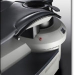
Advanced Research Technologies Inc. ("ART") announced the first sale of SoftScan® optical breast imaging system to the Sunnybrook Health Sciences Centre ('Sunnybrook") in Toronto, Canada. Sunnybrook is the first health centre to purchase a SoftScan imaging system since the Canadian company received regulatory approval for commercialization in Europe and Canada for its optical breast imaging…

The upper gastrointestinal (UGI) team at Milton Keynes Hospital NHS Foundation Trust has developed a rapid access service for upper gastrointestinal cancers. The new system means that patients are referred directly from their GP to the hospital for an endoscopy.
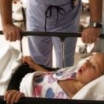
Evolving from an award-winning project carried out by undergraduates of the Technion-Israel Institute of Technology, Israel's leading University for science and technology, a new non-invasive device for detecting early stage respiratory irregularities is momentarily in development. The novel respiration monitor is intended to immediately detect deterioration in lung ventilation of ICU patients.
GE Healthcare recently acquired Image Diagnost International GmbH, an IT provider specialised on developing integrated software solutions for mammography workflow and image processing. With this acquisition GE Healthcare expands its capabilities in offering clinicians and national screening services an even more expanded portfolio for the detection of breast cancer.