Cardiac Diagnostic Imaging - Significance of ultrasound
Some of the medical procedures and equipment showcased and discussed at the MEDICA 2007 are sure to provide medical experts with plenty of discussion material. In cardiac diagnostic imaging a minor revolution is taking place, with ultrasound procedures increasing in significance. But innovation in x-ray methods is also continuing.
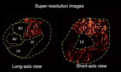
If the rapidly-beating heart can be scanned without camera shake, the same applies to the coronary vessels. This allows deposits in and strictures of the coronary arteries to be spatially represented for the first time. This could potentially enable computed tomography, a catheter-free imaging method, to replace traditional cardiac catheter examinations.One of the most important uses to which cardiologists are putting this new technology is in differentiating between the types of arteriosclerotic deposits in the coronary arteries - hard and calcified or soft and inflamed plaque. Four out of five heart attacks are triggered by soft plaque, which can detach and block a blood vessel to the heart. Distinguishing between the two prevents patients from undergoing unnecessary catheter therapy.
This could be made possible by another new CT concept in which two x-ray tubes rotate around the heart. These are operated with different levels of radiation, resulting in two images of the same region, each with a different tissue contrast. According to initial experiments performed by cardiologist Dr. Stephan Achenbach from the University Hospital of Erlangen, it may be possible to distinguish between less dangerous hard and extremely risky soft plaque.
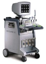
Another process, in which portable ultrasound equipment is used to perform cardiac evaluations, is becoming increasingly popular – particularly on seriously ill patients in intensive care. If a patient is too ill to be transported, a bedside ultrasound is almost the only diagnostic option. In such cases, however, it is crucial that an experienced doctor is on hand to interpret the ultrasound images correctly.
Transesophageal echocardiography (TEE) can be used to diagnose serious, previously undetected heart valve malfunctions, as well as pulmonary embolisms or myocarditis. The perennial issue is, of course, the identification of dangerous cardiac embolism sources.
“Echocardiography plays an important role when a patient is doing very poorly and you want to find out why that is as quickly as possible”, says Dr. Christian Bruch, a cardiologist at the University Hospital of Münster, explaining the TEE’s significance.
In two out of three unstable intensive care patients, the portable cardiac ultrasound equipment indicates the course of further treatment. The entire bedside echocardiographic evaluation is completed within 20 minutes.
03.09.2007