Hot Spots:
Nanoscale contrast agent for imaging coronary arteries
A next-generation diagnostic tool for cardiovascular disease, using a nanoscale iron particle, is now under development at a unique industry-government-university named Nano AG. A report from Siemens describes the research and progress at the centre
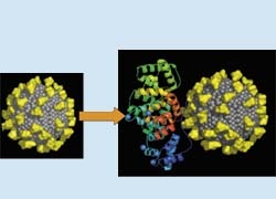
VSOP particle with yellow molecules indicating the citrate bound to the iron oxide surface (grey). Functionalisation of the particle by specific peptides e.g. Annexin V
A second application for the highly responsive nanoscale particles is more speculative, but could prove more valuable. With assistance from Siemens, the consortium leader of Nano AG, Prof. Taupitz and colleagues at other luminary academic sites are trying to attach the tiny particles to peptides that bind to specific structures in the body, which could turn the nanoparticles into disease–hunters inside the body.
To detect arterial plaque, the nanoscale iron oxide particles could be attached to a factor that would, in turn, attach to compounds involved in apoptosis. Programmed cell death in the artery wall is one sign of vulnerable plaque. A second tactic could link to compounds associated with angiogenesis, which often occurs in unstable plaque. In either case, the tiny particles will enrich within the pathologic vessel walls and generate hot spots on a MR image.
Currently, physicians must rely on indirect methods to specifically image diseased arteries. It is a change in paradigm for vascular diagnosis. We may have to look not so much at flow-limiting stenosis, or narrowing, but at the composition of the plaques and the change in the vessel walls. Using a specific contrast medium means to get functional information and then to make a prediction of the risk of plaque rupture in the artery.‘
01.09.2007
More on the subject: