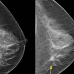
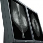
Photo

Radiation Treatment Planning

Radiotherapy treatment planning relies on transversal CT images. They form a basis for treatment planning, dose calculation and increasingly the plan localization for external radiation therapy, called CT simulation. A dedicated CT scanner for radiotherapy plan simulation is one of the most essential pieces of equipment in a modern radiotherapy department. In contrast to external beam…

Photo

Smooth workflow, excellent diagnostics and patient-friendly handling

With 715 beds, the University Hospital Maastricht is one of the leading hospitals in the Netherlands. The modern building offers 22 operating theatres and in 2005 24,362 patients were admitted. Due to rotating staff systems and multidisciplinary responsibilities in diagnostic imaging departments in large hospitals like the University Hospital Maastricht, the workflow has to be as easy as possible…

Photo

Clinical Experience with a Superconducting MRI Scanner

Following the installation of a 0.5-T superconducting magnetic resonance imaging (MRI) system at our hospital in 1987, we have been performing a large number of examinations as a key regional hospital serving the city of Muroran, Hokkaido, and surrounding areas. However, compared with more recently developed MRI systems, 0.5-T systems tend to have poorer image quality and a restricted range of…

Photo

Sonographic Indications of Fetal Chromosome Abnormalities

"Genetic ultrasound" is a refined technique in prenatal diagnostics. It aims to estimate as accurately as possible and non-invasively, the individual risk of a fetal chromosome abnormality. In conventional prenatal sonography (malformation screening) conspicuous fetal malformations are searched for selectively; naturally chromosomal anomalies of the fetus cannot be detected in this…

Photo

Tissue Doppler Imaging

Tissue Doppler Imaging (TDI) is an emergingnon-invasive ultrasound technique that makes it possible to measure velocities at any point of the ventricular wall during the cardiac cycle. Over the last ten years, TDI has been proposed as a feasible and useful imaging tool that provides information on regional wall motion dynamics.

Photo

Selenium-Based Flat Panel X-ray Detector for Digital Fluoroscopy and Radiography

We have succeeded in developing the first selenium-based flat panel X-ray detector for digital fluoroscopy and radiography. This flat panel X-ray detector efficiently captures X-rays that have passed through the patient's body and converts them directly to digital dynamic and static images. It has been confirmed that images with high spatial resolution and contrast can be obtained. The detector…

Photo

Enviromental Protection in medical technology

For the production of medical technology products more and more companies take enviromental protection and energy efficiency into consideration. Siemens Medical Solutions e.g. ensures with a holistic concept that the products not only match high ecological demands during production, but also in operation and beyond shutdown.

Photo

Endoscopic Ultrasound Transducer with FNA Biopsy Capability

Endoscopic Ultrasound with Fine Needle Aspiration Biopsy (FNAB) capability is relatively new for Toshiba. Recently the company has introduced a transducer enabling gastroenterologists to perform these kinds of examinations. Dr. Paul Fockens (PhD), Director of Endoscopy at the Academic Medical Centre (AMC) of the University of Amsterdam, is an expert in this field. He has been Director of…

Photo

Technical Insights into the Dual-slice CT

In 1999, the Aquillion/MULTI and Asteion/MULTI multi-slice systems with highly integrated 34-channel detector system, both consisting of a total of 30,464 detector elements, were introduced. On the basis of such a structured detector matrix, a new image reconstruction procedure was used. This so called MUSCOT (Multislice Conebeam Tomography) procedure made the dream of an isotropic voxel in the…

Photo

Multi-slice Helical CT Scanning of the Chest

Lung cancer is the main cause of deaths due to cancer in human males and the incidence is constantly increasing. A cure rate of only 10% has been reported because most neoplastic lesions are diagnosed too late. However, when lung cancer is diagnosed early and surgical resection performed (stage 1 cancer), 5-year survival is significantly improved and may reach 70%.

Photo

Sure Start

The use of state-of-the-art helical CT scanners allows for ultra fast examination of larger regions of the body. Due to the short examination time, optimum utilization of the intravenous contrast medium bolus is of extreme importance. The Sure Start function grants this in a very simple way.

Photo

Cardiac CT

Along with a high local resolution, the cardiac CT imaging also requires a high time resolution where the image data acquisition must be synchronised with the cardiac cycle by means of parallel ECG recording, and the measuring volume must be acquired in a single breathhold cycle. The multi-slice CT copes well with these requirements as it allows for acquiring continuous volume data sets of the…

Photo

X-Rayed Masterpieces

Even today, 105 years later, the X-rays have not lost their fascination and importance. The discovery of X-rays in November 1895 by the physicist Wilhelm Conrad Roentgen introduced the 'X-ray era' and caused great change in all areas of life. One of the most fascinating fields of research is the analysis of works of art by means of X-rays. The 'anatomical structure', i.e. the composition of the…

Photo

Magic Microbubbles

The introduction of a new generation of microbubbles has created opportunities for ultrasound, and this is particularly true in the liver, where study of the arterial, venous and sinusoidal phases with an agent such as SonoVue at low ultrasound power levels (Iow mechanical index, MI) provides unique information. Similar diagnostic advantages can be obtained in diseases of the kidneys, spleen and…

Photo

Learning from the Human Brain

In today's high end diagnostics and clinical research ultrasound systems are required that combine outstanding performance with excellent ease of use. With APLlO, Toshiba has developed a new platform that meets the highest demands for diagnostic confidence and workflow optimization. It is developed based on a groundbreaking new system architecture. This has made the implementation of numerous…

Photo

Aplio - Dynamic and Advanced Dynamic Flow

There are a number of problems in contrast imaging using LevovistTM contrast agent. In Doppler mode, the problems are poor resolution and large areas of blooming. In 2nd harmonic imaging and pulse inversion imaging, there are problems related to tissue harmonic imaging (THI) because Levovist requires a high mechanical index (MI). THI increases under high-MI conditions, and THI interferes with…

Photo

Upper Gastrointestinal Tract Endoscopic Ultrasound

Endoscopic ultrasound (EUS) is the combination of ultrasound and endoscopy, which allows placement of high-frequency transducers close to the intestinal wall and adjacent structures. As EUS enters its third decade, it is used for locoregional staging of many upper gastrointestinal malignancies, including cancers of the oesophagus, stomach, duodenum, ampulla of Vater, extrahepatic bile ducts and…

Photo

Automatic Quantification of Left-ventricular Function

The evaluation of the size and function of the left ventricle in patients with suspected heart disease is a central diagnostic problem. In contrast to other methods of evaluating left-ventricular function (e.g., angiocardiography, right-sided heart catheterization, radionuclide ventriculography or MRI), transthoracic echocardiography is a widespread and readily available procedure that is not…

Photo

High Resolution Protocol Optimization

Since the introduction of multi-slice computerized tomography (MS-CT), several authors have reported an increased exposure to radiation when using the manufacturer-specific protocols compared with the previous technique of single-slice computerized tomography (SS-CT). On the basis of comprehensive dose measurements conducted on a phantom, a study by S. M. Giacomuzzi et al reported that the mean…

Photo

Procedure and Clinical Value of CT Fluoroscopy

The past several years, interventions using ultrasound for real-time image monitoring in diagnostic and therapeutic procedures have increasingly been integrated into the daily clinical routine. Limitations due to image-distorting physical effects, such as the total reflection at the soft tissue/lung boundary, are associated with the use of ultrasound. CT fluoroscopy on the other hand enables…

Photo

Lasting for a day - Ephemera in radiology

There has been a popular fascination with X-rays ever since their discovery by Wilhelm Conrad Röntgen. And this fascination shows no signs of abating. Much of the material that is of interest for the historian of radiology is transient. The term "ephemera" covers a large range of material including leaflets, trade cards and tickets. This type of material has not traditionally been kept…

Photo

Multi-slice CT: Learning from experience

The Netherlands - September '00 - A good example of one of the discussions was the general issue of dose reduction. All participants agreed that it would be a challenge to be able to realise this, although this should definitely not interfere with the image quality. An interesting challenge for the users as well as the manufacturers! Many interesting topics were presented during the seminar, of…

Photo

Tissue Doppler Imaging for assessment of pseudonormalization of the mitral inflow pattern

Impairment of left ventricular relaxation may lead to pulmonary congestion and symptoms of heart failure even in patients with preserved systolic function. Doppler echocardiography has become the non-invasive method of choice for the assessment of both systolic and diastolic dysfunction. However, Doppler-derived parameters such as mitral inflow velocity, deceleration time and isovolumic…

Photo

Contrast Harmonic Imaging (CHI)

Case 1 50-year-old male with a colorectal cancer. Greyscale imaging clearly shows the lesion, as an echogenic 3 cm mass (Fig.l a), which must be considered as suspicious for a metastasis from his colorectal cancer but benefit of the doubt must be given. Colour Doppler (Fig. 1b) is not helpful. At 33 seconds after SonoVue injection (early sinusoidal phase), Vascular Recognition Imaging (VRI)…

342 show more articles
Subscribe to Newsletter