
euHeart
Royal Philips Electronics is to lead `euHeart´, the new European Union (EU) funded research project that aims to improve CVD diagnosis, therapy planning and treatment.


Royal Philips Electronics is to lead `euHeart´, the new European Union (EU) funded research project that aims to improve CVD diagnosis, therapy planning and treatment.

Every summer the European Society of Cardiology (ESC) holds Europe's biggest annual meeting of specialists in cardiovascular medicine, inviting and drawing in top international medical professionals. Karoline Laarmann asked Professor Kim Fox, President of the European Society of Cardiology and Consultant Cardiologist at the Royal Brompton Hospital, and professor of clinical cardiology at Imperial…

More than 4,500 cancer and radiotherapy specialists will gather in Göteborg, Sweden, this September for the 27th annual congress of ESTRO. ESTRO 27 is to be held between the 14th and 18th September 2008 at the Göteborg Convention Centre in Göteborg.
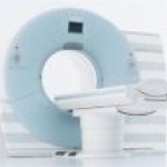
A new study reveals that, with dual-source computed tomography (DSCT), the effective dosage for a heart examination can be significantly lowered, in comparison to conventional computed tomography (CT). The study also demonstrated that stenoses can be diagnosed with the same high accuracy as with invasive x-ray angiography.

US- researchers found that inpatients experience an average of nearly one and a half potentially harmful errors in their medication record during a hospital stay. They started the search for the causes: admission and discharging are the most dangerous situations.
Women who survive breast cancer for at least five years have a 89% chance that it will not recure. But those, who are still suffering and on endocrine treatment are likely to get arthralgia and arthritis.

The “Project Connect”, a program designed to build Public Private Partnerships to combat HIV and tuberculosis in India recently recorded a huge success:
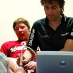
Compact ultrasound technology provides greater diagnostic confidence within close proximity of athletes. The portable equipment from GE Healthcare is not much larger than a laptop, and delivers comparable results in terms of display quality to the high-end equipment used in hospitals.
More than 250,000 women under the age of forty are living with the disease in the US and 11,000 will be diagnosed in the next year. Even so, young women are underrepresented in many research studies and treatments, according to genomic expert Simon Chin.
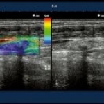
The idea of measuring tissue stiffness using ultrasound is nothing new. But lately the field sets a rapid pace. “Real-time, hand-held elastography is now a commercial reality,” asserts Jonathan Ophir from the University of Texas Medical School.
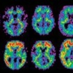
The so-called 11-labeled Pittsburgh Compound B ([11C]PiB) type of positron emission tomography (PET) may be useful in a non-invasive assessment of the formation of Alzheimer's disease-related plaques, according to a study that will appear in the October 2008 issue of Archives of Neurology.

To make sure that for athletes and officials the Beijing 2008 Olympic Games will not come to a premature end, digital medical and dental imaging systems from Carestream Health, Inc. are available to diagnose and treat any health concerns.
Treadmill exercise testing is a common tool to detect of cardiovascular diseases. But clear images of the working heart are hard to obtain. Now researchers designed MRI equipment to provide high-resolution images of the heart at critical stages.
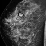
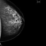
Researchers compared the Norwegian organised population based mammogram screening every second year and a physician- or self-referrals annual test in the US. Both are equally sensitive, but the recall rate for abnormal results was lower in Norway.
Tiny lipid-shelled microbubbles injected in vessels may serve as an new ultrasound contrast agent to evaluate microvascular blood flow. By this microbubbles US-researcher devised a ultrasound imaging technique that can spot early signs of PAD.

The 10/66 Dementia Research Group warns that dementia is often not diagnosed in low-income and middle-income countries. Latest research shows that these figures has been underestimated and the economic costs of dementia and other age-related illnesses are rising.

This May the Brussels-based Crossroads Institute for Cardiac and Vascular Medical Education launched two new educational courses on the prevention of amputation (peripheral vascular disease) and on improving the treatment of women with cardiovascular disease.

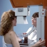
It sounds impressively simple: An over-the-counter pain-relieving gel from the drugstore may reduce the uncomfortable and even dolorous feeling women experience during mammography exams, according to the a study published in the online edition of Radiology.

Totoku now offers the new 19inch, 1,3 Megapixel grayscale display ME191L. It supports all modality devices of the most common manufacturers but fits also for special applications like cardiology. The display was developed to replace the CRT monitors, which reached the end of their lifetime.

A new type of super-resolution X-ray microscope invented by researchers from Paul Scherrer Institut (PSI) and Ecole Polytechnique Fédérale de Lausanne (EPFL) gives pin sharped insights into the composition of semiconductor devices and cellular structures.

For the second time the European Centre for Disease Prevention and Control (ECDC) in Stockholm, Sweden, invites to the European Scientific Conference on Applied Infectious Disease Epidemiology (ESCAIDE). The ECDC published now a forecast about expectations and the global hot topics of the international event in Berlin, November 19-21.
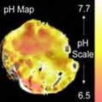
Diseases are often associated with a low tissue pH. Researchers from the UK and Sweden have now developed a MR imaging method to measure the pH in human body using 'baking soda'. The procedure might display the pathological process of inflammation or cancer, as well as the response to the therapy.

iTunes can more than only play music, according to a new study. The successful music-managing software from Apple allows its user to manage and organize PDF files just as easy as a record collection, making it possible for radiologists to better organize their personal files of articles and images.
GE Healthcare today announced John Dineen as the companies new CEO and president. Therewith Dineen changes from the Transportation to the Medical Technologies and Services Segment of General Electric.

With the help of an engineered common cold virus spreadings of prostate cancer in the pelvic lymph nodes can be visualised with a PET scanner. It is now possible to treat the cancer in an early stage. But the developers of the virus from UCLA's Jonsson Comprehensive Cancer Center see a chance to even use it as a treatment option.