Real-time Tissue Elastography
Aiming for a `manogram´programme in Europe
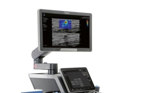
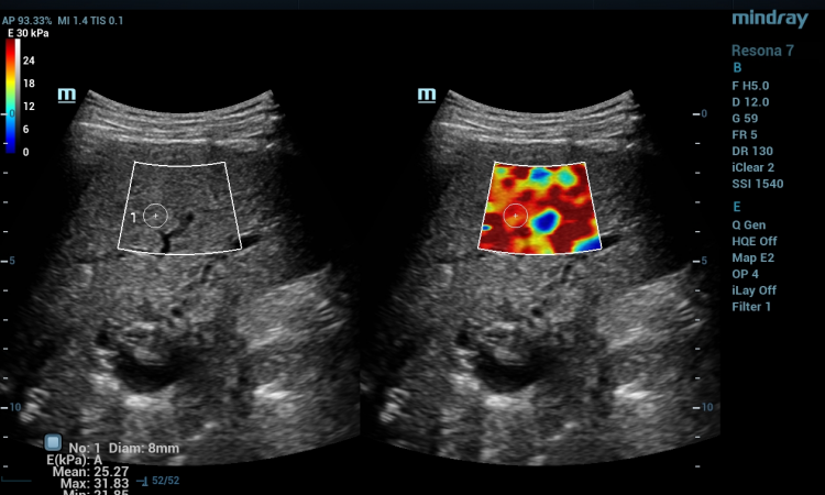
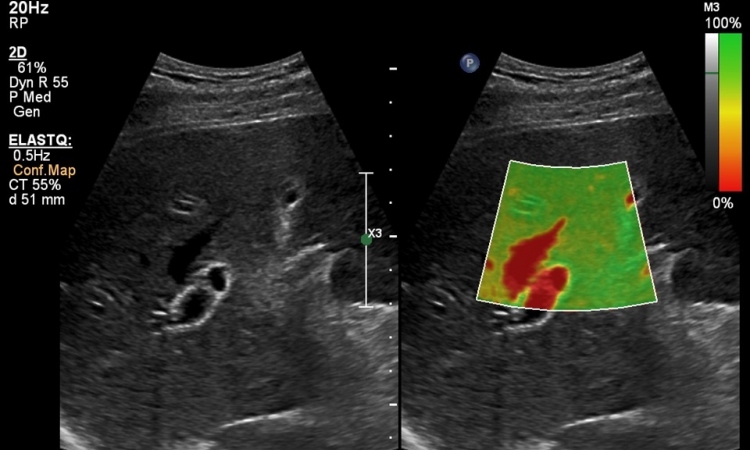
Today, Real-time Tissue Elastography (HI-RTE) provides a method to determine tissue elasticity of certain organs, such as the prostate, in real-time and to perform precise biopsies for reliable tumour diagnosis during standard examinations.
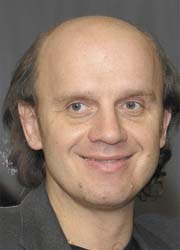
Professor Ferdinand Frauscher (FF), consultant in radio diagnostics at the Innsbruck University Hospital, Austria, who has pioneered elastography, particularly in urogenital diagnostics, and regularly uses it in a screening programme, discussed the value of this method in an interview with Meike Lerner
FF: Real-time elastography is gaining increasing importance, because it is a patient-friendly, reliable and very affordable method to diagnose cancer. The idea behind the method – to measure elasticity and thus density of tissue – is not new. However, for a long time this kind of measurement required complex and time-intensive procedures and therefore was not useful in clinical practice. The introduction of HI-RTE by Hitachi Medical Systems offered a rather simple method which can be applied routinely.
HI-RTE has become an invaluable tool particularly for the diagnosis of prostate cancer, because, before this, systemic (‘blind’) biopsy had been the only method. In Innsbruck we have been running a prostate cancer early detection programme with PSA (prostate-specific antigen) screening since 1988. Within this programme we examined many men who did not present with complaints, but where a prostate carcinoma was suspected. Before the introduction of real-time elastography we had to perform a systemic biopsy, which in 40% of all cases generates false negative results. From a radiological viewpoint there was an urgent need for an imaging method that allows visualisation of the prostate. Around 1993, we started to work with colour Doppler ultrasound, which we used until real-time elastography became an alternative.
Today, we have data on about 1,500 patients for whom we used real-time elastography to visualise known prostate cancer. Before radical prostatectomy the patients were examined with elastography, the tissue density was recorded and results were compared with the post-surgical results. Sensitivity of the method reached 90%, specificity around 70%.
We also compared real-time elastography with MRI. In screening programmes MRI is significantly more time and cost intensive and more unpleasant for the patient. An examination with MR-guided biopsy takes up to three hours - that is unfeasible in every-day clinical practice, because only two to three patients can be examined per day.
The Federal State of Tyrol is one of few regions that offer a comprehensive prostate screening programme. What kind of patients present?
FF: Most of our patients show a PSA value between four and five. Here, in Tyrol, we set the screening threshold at 1.25 since earlier studies indicated that in 20% of men with a PSA value below four a tumour was detected. That means even a slight increase in PSA may indicate cancer. On the other hand, a harmless infection can also cause an increased value. With real-time elastography we can perform a transparent examination that is painless for the patient. Combined with a biopsy the detection rate reaches 60-70%.
Real-time elastography is also excellently suited for therapy control. Patients with dense tissue are scheduled for regular follow-up exams, as are patients with a low-aggressive tumour that does not receive treatment.
Today, real-time elastography is being used in two further Austrian clinics and in a hospital in Hamburg, Germany.
So, thanks to real-time elastography the diagnosis of prostate cancer has been significantly improved. What are the next steps?
FF: Next, we want to look at possible new therapies. As far as men are concerned, we are lagging behind a bit. Our goal is – similar to breast cancer therapy – to avoid radical resection if at all possible. Resection is still the procedure of choice, even if a tumour has a size of only three to four millimetres. With the support of imaging methods, we can treat these cases with high-focus ultrasound or can apply special gene therapies. The US is ahead of us, because more funds are available for research. The term ‘manogram’ is already widely used in the US. We would like to adapt this trend for Europe, which would result in significantly improved diagnostics and treatment of prostate cancer.
Real-time Tissue Elastography at the ECR
Organised by Hitachi Medical Systems, the symposium ‘Real-time Tissue Elastography: stretching diagnostic boundaries – myth or valuable clinical tool?’ will be held during the European Congress of Radiology (ECR), on Sunday, 9 March (12:30-13:30, room E1).
High profile specialists will present the latest developments in RTE, and Prof. Frauscher will describe his experiences with this method and explain ‘The value of real-time elastography in the diagnostic evaluation of prostate cancer and testicular masses’.
01.03.2008