CAD improves breast cancer diagnosis
A study recently published in the online version of the American Journal of Roentgenology evaluated the clinical value of Computer-Aided Detection (CAD) software in the interpretation of mammograms. In the course of the study, the recall rate, sensitivity, positive predictive value, and cancer detection rate for single reading with CAD, versus double reading without CAD, were investigated.
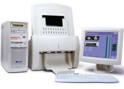
In his study, which also compared Biopsy and pathology data for positive cases, Dr. Matthew Gromet of the Breast Imaging Section of Charlotte Radiology, Charlotte, USA, found that mammogramms read by a single radiologists with the aid of CAD had a statistically significant increase in sensitivity (11%) and a smaller increase in recall rate (4%) than mammograms read by a single radiologist without CAD review.
Even when compared with independent double reading without CAD, which is the standard in European breast cancer screening programmes, the single reader CAD reviewed results proved near equal in sensitivity, but showed a statistically significant lower recall rate.
Dr. Gromet’s study results base on the performance of nine radiologists from Charlotte Radiology with a mean level of experience in mammography of 15 years. The radiologists interpreted a total number of 231,221 screen-film Mammograms, which were obtained on Hologic mammograpgy equipment. 49% of the exams were double read without CAD and 51% were single read with the Hologic R2 ImageChecker® CAD system. “CAD appears to be an effective alternative that provides similar, and potentially greater, benefits,” comments Dr. Gromet. His results support the clinical practice of CAD becoming increasingly popular as an alternative way to increase sensitivity, since double reading is very time-consuming.
“The Gromet study is a valuable addition to the body of knowledge on the performance of radiologists reading screening mammograms with CAD assistance,” adds Ronald A. Castellino, MD, FACR, and Chief Medical Officer for Hologic.
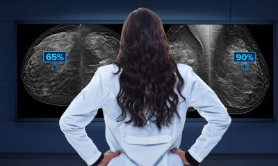
His company sees great potential in CAD, that marks the areas of interest in mammogramms that might otherwise be overlooked by interpreting radiologists. However, the software also tends to place false marks, identifying areas of suspicion that are not cancer. Eventually the radiologist is still responsible for the interpretation of an examination and identication of lesions.
29.02.2008


