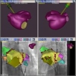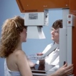
Apple gets ads from radiologists
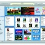
iTunes can more than only play music, according to a new study. The successful music-managing software from Apple allows its user to manage and organize PDF files just as easy as a record collection, making it possible for radiologists to better organize their personal files of articles and images.