News • Imaging equipment
GE Healthcare is presenting ways to see more
At RSNA 2009, GE Healthcare is presenting a big basket filled with trends and innovations for the radiology department:


At RSNA 2009, GE Healthcare is presenting a big basket filled with trends and innovations for the radiology department:
Two members of the Heart Center at the University of Leipzig teamed up during Medica for a tour de force presentation on Future Trends in Cardiac Surgery. "The aim of the game is opening the chest through little keyholes to operate in the most minimally invasive way possible and avoid sternotomy," said Prof. Friedrich Mohr, Program Director at the Leipzig Heart Center, who review new surgical…
Sonoace, the German distributor of the Korean firm Medison Co. Ltd has launched its first hand carried ultrasound machine MySono U5 at Medica. ‘Complete with various image optimisation processing technologies in a lightweight and compact form , Mysono U5 is designed to guarantee immediate diagnostics in a wide range of applications, such as emergency medicine, vascular, musculoskeletal and…
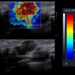
French expert Dr Jacques Souquet PhD, President of SuperSonic Imagine, describes the value and potential of ShearWave elastography, the latest development in ultrasound that enables radiologists to acquire user independent information about tissue stiffness by measuring both, ultrasound and shear waves.
In recent years Hitachi Medical, the pioneer in developing ultrasound elastography to diagnose tissue stiffness, has seen competing systems launched that claim to have the same capability. ‘We are approaching a point where elastography will be considered a standard feature, call it the E-mode, for ultrasound,’ Heinz Schreiber, head of Ultrasound for Hitachi Medical Systems Europe told…
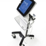
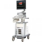
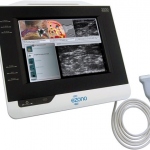
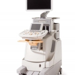
GE Healthcare is presenting its new Venue ultrasound product line at Medica. The Venue 40, the first product launched, provides visualisation for needle guidance procedures and rapid diagnostics in real-time at the bedside. These point-of-care (POC) settings are the fastest growing in ultrasound internationally (USA growth: 30% average in the last four years. Source: industry report by Klein…
Ultrasound presents a promising technology for non-invasive examinations of the abdomen. There is a vast, unmet medical need in the area of liver fibrosis for the clinical assessment of patients with fibrosis who either refuse or cannot undergo the painful and invasive biopsy procedures that provide hard histological evidence of the state of the disease.
Mirror 2, the all-digital colour Doppler ultrasound system, produced by Landwind International Medical Science Pte Ltd, incorporates a number of advanced imaging technologies, including powerful multi-beam parallel imaging, premium vascular imaging, real-time dynamic receiving focusing, magic focus, superior aptitude filter, 3-D, and panoramic imaging, the manufacturer reports.
Following the October commencement of shipments of its new, compact, mobile, colour ultrasound system ProSound Alpha 6, the manufacturer Aloka Holding Europe AG has brought the system for demonstrations at Medica.
A portable system designed for anaesthetists, the eZono accelerates the learning curve with the manufacturer’s patented Cue Cards, a multi-media learning and workflow tool for vascular access and regional anaesthesia - combined with excellent image quality, the manufacturer points out.
Founded in Poland, in 1993, Echo-Son S.A. manufactures the ultrasound colour Doppler scanners Spinel II, Epidot_SC, Desmin_M, Epidot_V and the portable (b/w) Desmin_F, Desmin_H and Albit, for medical and veterinary applications (2.5-12.0 MHz, Doppler) and ophthalmology: A+B mode scanner 12 MHz and pachymeter 20MHz.

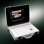
Specially designed for mobile general practitioners and urologists, the Draminski Ultrasound Scanner is fully portable and comes in a solid and secure case that also includes all necessary accessories for diagnoses.
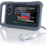
The S8, a high-end colour Doppler HCU (hand-carried ultrasound) recently received the 2009 Product Quality Leadership Award from the global consultancy Frost & Sullivan (F&S).
Sonoace, the German distributor for Medison Co. Ltd, has announced the launch of a new, dedicated high performance OB/GYN ultrasound system, the ACCUVIX V20 ‘Prestige’, which features the industry's first multi-rendering 3-D/4-D technology, 3-D MXI (3-D Multi-eXtended Imaging ).
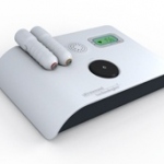
Weighing just 2 KG, in a hospital, small clinic or doctor’s surgery the fully portable Fetatrack DD250 can provide continuous foetal and vascular dopler. The unit operates from a mains electricity supply for 30 hours; alternatively the built-in rechargable batteries can be used. ‘The system has all the necessary facilities for accurate antenatal foetal assessment in one compact unit,’…
‘Supremely portable and incredibly tough, the NanoMaxx ultrasound system combines Best in Class performance with affordability and simplicity,’ the Erlangen-based manufacturer SonoSite reports.
Designed for multi-applications (e.g. general imaging, obstetrics/gynaecology, abdomen, small parts, musculoskeletal, urology, vascular) MyLab 20Plus, manufactured by Esaote Biomedica Deutschland GmbH, can be customised to suit individual needs in a shared ultrasound department or clinic.
Philips has developed a proprietary technology for elastography that uses new strain analytics for signals captured on the IU22 volume linear transducer and yields a relative quantification of tissue stiffness, which is presented as statistical charts displayed alongside a colour-coded qualificative image superimposed on the B-mode screen.

The University of Pennsylvania Health System, one of the largest health systems in the US, has awarded a $135 million contract for Integrated Service Management to Siemens Healthcare. Over the next seven years, Siemens will be servicing and ensuring around-the-clock availability of diagnostic systems and bio-medical devices from diverse manufacturers in the customer´s various hospitals.

Emphasising the crucial partnership of radiologists and urologists in the treatment of prostate cancer, Professor Arnulf Stenzl, Medical Director of Urology at the Tübingen University Hospital, explained that, throughout the phases of prostate cancer diagnosis and therapy - from primary diagnosis onward - imaging is indispensable.

During a gathering of clinicians, scientists and economists at this year's Medical Technology Congress (Treffpunkt für Medizintechnik) held in Berlin's Charité Hospital, all 17 lectures focused on cancer treatments. Bettina Döbereiner reports
The respiratory unit at Newcastle's Royal Victoria Infirmary (RVI) is using SonoSite's M-Turbo system for management of pleural effusions, particularly in patients with lung tumours.

Professor Wolfgang Heinrichs, of the AQAI Simulation Centre Mainz, Germany, notes that the 15th SESAM congress slogan "Europe-tradition and innovation in simulation", was born out by the choice of venue, the Mainz Simulation Centre was one of the first of its kind in Europe, and also hosted the 2000 SESAM congress.
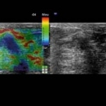
Real-time Tissue Elastography, an ultrasound procedure that uses colour to differentiate clearly and unambiguously between malignant, hardened tissue and flexible, benign tissue could be the addition needed to have really effective breast cancer screening, according to independent radiologist, and specialist in Real-time Tissue Elastography Dr Bruno Scheffer from Nantes, France.