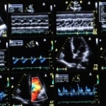
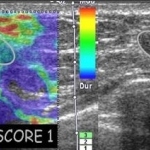
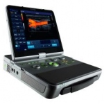
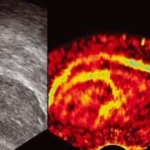
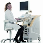
Ultrasound

Photo

Scanning children

A growing number of Dutch hospital doctors now favour ultrasound (US) over CT scans to detect appendicitis in children. This trend has re-opened the US v. CT debate over which diagnostic method is more effective, cost-effective and safest for a child's longer-term health needs.

Photo

LOGIQ E9

GE Healthcare in unveiling LOGIQ E9, a pioneering sonography system that can generate a precise image fusion from CT/MRT and sonographic volume data, the firm reports.

Photo

HI VISION 900

Hitachi has developed a new abdominal sonography system, based on a fully digital platform with a unique high-performance computer and an innovative octal parallel wideband beamformer, the firm reports.

Photo

Elastoscan in mammary sonography

If, in the early days of mammary sonography, it was revealed to be helpful to render tactual findings in a visible manner (sonic palpation), then now is the time to palpate clinically occult findings in a new manner (Elastoscan).

Sonoelastography makes hardened cancer tissue visible

Sonoelastography, a procedure that measures the elasticity of tissue and differentiates between healthy and hardened pathological tissue, will make cancer diagnosis safer in the future. For example, it has been shown to improve the accuracy of prostate cancer diagnosis by 20%.

Photo

Ruhr's centre of excellence for medical

The Centre for Medical Imaging Ruhr (zmb ruhr) in Bochum offers the technological infrastructure for the development of therapeutic and diagnostic imaging procedures. The primary focus is on ultrasound — the most commonly used imaging procedure in Germany, delivering the highest volume of examinations.

Photo

ACUSON S2000 with Virtual Touch improves liver diagnosis

Siemens Healthcare is showing a very innovative application on the new ACUSON S2000 ultrasound system. “The software packages Virtual Touch tissue imaging and Virtual Touch tissue quantification represent the clinical realisation of a method, previously known as Acoustic Radiation Force Impulse (ARFI) technology”, the firm explains.

Photo

First OCT tissue images

A UK collaboration, led by an optical imaging company has produced its first optical coherence tomography (OCT) tissue images at a wavelength of 1 µm. Based on interferometry, OCT works by analysing near-infrared light reflected from a sample and using differences in the reflected intensities to create high-resolution images. Employing 1 µm light - as opposed to the longer wavelength…

Photo

C-Arm technology in daily use

Last year, orthopaedics and sports medicine specialist Dr Rainer Burgkart (left), who is also a musculoskeletal researcher, selected a new C-Arm for use at the Technical University of Munich (Klinikum rechts der Isar). Recently, we asked him for the reasons behind this choice and his subsequent experience with this device.

Photo

The Carestream DRX-1

This September, Carestream Health revealed the first wireless cassette-size DR Detector. The Carestream DRX-1 system, which incorporates a console and wireless 14x17 inch cassette-size digital radiography (DR) detector, provides a rapid, affordable conversion for users of radiographic film or computed radiography systems, Carestream explains.

Photo

First wireless DR detector

Carestream Health's engineers created the first wireless DR detector which is not bigger than a standard cassette and can therefore withstand the challenging environment of a modern x-ray department. The rugged detector, case, and internal components are made to produce DR images of high quality under real-life x-ray department conditions.

Photo

Sono-elastography

Martini Clinic, in the University Hospital Eppendorf (UKE) campus in Hamburg, is one of the world's major centres for nerve-sparing prostatectomy. The Martini physicians not only specialise in surgical intervention but are also at the cutting edge of diagnostics, being among the few in Germany to use sono-elastography, an innovative tool to detect prostate cancer. We asked Dr Georg Salomon (GS),…

New Molecular Imaging Techniques Aim at Detection of Earliest Steps of Disease Development

An emerging discipline of noninvasive cardiac imaging, molecular imaging, has evolved constantly in the last few years and is increasingly being translated from the preclinical to the clinical level. Molecular imaging allows for unique insights into specific disease mechanisms and holds great promise to change the practice of cardiovascular medicine by facilitating early disease detection,…

157 show more articles
Subscribe to Newsletter