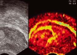
Sonoelastography´s 2nd generation tools aid research
The early, safe detection of prostate cancer is a challenge.
Current procedures are still imprecise and speculative; without a subsequent biopsy suspicious PSA levels, images and the results of palpation cannot be correctly evaluated.
Thus there is an energetic pursuit to discover new imaging technologies. Dr Thilo Eggert at the Urological Clinic of the Ruhr University Bochum at the Marienhopital Herne, is among the investigators.

‘There are different research approaches to optimise early detection,’ he explained. These include research into new serum markers ‘to more clearly specify the PSA level, because this is currently our only laboratory parameter for suspected cancer’. There are also different approaches involving imaging to achieve earlier and more accurate detection of carcinoma. ‘The elasticity or cell density of tumour tissue gives us particularly valuable information here,’ he pointed out. In this, he rates sonoelastography as a promising approach to early detection of carcinoma. Although clinical researchers have pinned hopes on this procedure for some time, during initial trials its technology was not fully developed and perfected sufficiently. ‘The first generation sonoelastography equipment fell short of expectations, compared with B- image sonography. It was then not possible to confirm a significant improvement in the early detection of prostate cancer through sonoelastography-supported biopsy studies,’ Dr Eggert said. ‘However, the latest generation of equipment from Hitachi, which features vital, up-to-date technologies, convinces.
The handling, as well as sensitive imaging of tissue hardness through colour scales, has been vastly improved through the new technology.’
Another new feature of this equipment is that the user is shown how much pressure needs to be exerted on the tissue to produce constant sonoelastography images. This reduces the respective dependency on who is carrying out an examination and produces a basis for reproducible results. The opportunity to combine sonoelastographic image information with conventional B-images makes it easier for an examiner to carry out well-targeted biopsies.
Sonoelastography is thus experiencing its first Renaissance. We need new studies to achieve clear evidence about the benefit of this diagnostic tool.
Dr Eggert is currently using the Hitachi equipment for examinations. ‘We will compare the data gained through sonoelastography of patients with confirmed cancer with the histological result of a prostatectomy preparation prior to surgery, to test the sensitivity and specificity compared with conventional ultrasound. In another part of the study we are testing the system for initial biopsies on patients with suspected prostate cancer.’ To achieve truly meaningful data, he said the study cohort will involve around 600 patients so study results will not be available until 2009. ‘Until then, tissue measuring should only be viewed as a complementary procedure.’
01.03.2009


