Breast Diagnostics
Automated ultrasound delivers the first volume images of the breast
The diagnostic use of ultrasound has improved significantly because of improved resolution and reduction in artifacts, allowing increasingly more specific examination.

D Uhlenbrock
Further progress in breast diagnostics has now been made in the shape of volume scanning, which facilitates a 3-D visualisation of the entire breast, along with an efficient analysis of the data and semi-automatic reporting.
Professor D Uhlenbrock (DU) of the Medical Centre for Radiology and Nuclear Medicine at the St. Josefs Hospital in Dortmund, Germany, is among the first to use the Automated Breast Volume Scanner (ABVS) to diagnose breast cancer. In an interview with Daniela Zimmerman, he outlined the current and future use of this technology.
Having worked with the Acuson S 2000 Automated Breast Volume Scanner for just under three months, what differences have you found between this and conventional ultrasound scanners?
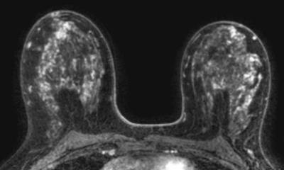
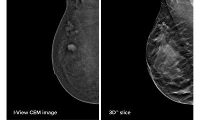
DU: ‘The Acuson S is basically a normal ultrasound system that is hand-guided and can be used for all ultrasound scanning indications. However, its particular feature is a 15cm scanning unit, the transducer, which can be used to create a 15x17x6 cm data set. The receptor is guided over the breast and delivers a volume image of the breast – similar to tomosynthesis. This data capture lasts between 60 and 90 seconds and between 250 and 400 images are taken during this time. The 2-D set of data is then transformed into a 3-D image. And this is the revolutionary feature: This type of examination is not possible with a 5cm transducer, because it would have to be used in a step-by-step way. Moreover, images gathered in this way previously couldn’t be saved in this form.
What are its advantages in daily practice?
‘The critical difference is that you can look at and evaluate a complete set of data of the entire breast at any time. A further advantage is automation; the pre-settings allow image acquisition to be carried out by a radiographer, because the diagnosis can be carried out by the doctor at a later date. It’s also very interesting for practical use that the patient lies on her back during the examination – just as she would later, during surgery. This means the data gathered precisely reflect the picture that would present itself to the surgeon. This means that marker points can be set and tumours localised at an early stage. And finally, you can actually switch the equipment to the volume ultrasound setting from the normal ultrasound setting at the touch of a button. This is quick and easy and extremely user-friendly.
Are there also limitations or disadvantages in this technology?
‘We are concerned about certain things. However, it remains to be seen whether they will actually turn out to be disadvantages. As this procedure is still very new we can, for example, not yet say precisely how the images can be evaluated in the best possible way. We receive 150 to 300 images in one set of data.
So one of the first questions are: Do I have to look at all of them? What level of slice thickness do I have to look at?
‘We must establish how much expenditure of time the post-processing and analysis of the images entails. We don’t yet have any valid results from studies on this. The question that then follows is: What is the diagnostic use versus the amount of time spent? I can say from experience that one aspect of this use is that we get an image of the entire breast and can be sure that no area has been missed.
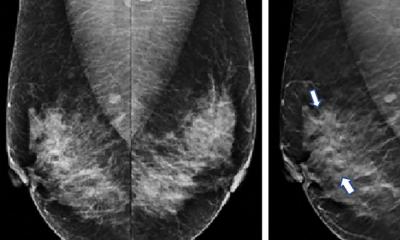
‘Finally, the last question is: Is the detection rate just as good? We have the first results from patients who had a clear diagnosis during mammography and who we then scanned with the Acuson S 2000. The result was that lesions smaller than 5mm were harder to detect. One reason for this is certainly that the size of the stored set of data tempts us just to quickly scan across all the images, and that important information may be missed. ‘However, once the procedure is established and we have sufficient parameters then a well-functioning CAD system can close this gap.
‘In my personal opinion, ABVS will become part of standard breast diagnostics in a few years time. The general trend in ultrasound scanning is heading towards a situation where the actual examinations will no longer primarily be the responsibility of doctors. Examinations will be read on a workstation by doctors after the scans have been taken. Then, we have to wait and see where exactly in the chain of diagnosis the procedure will find its place. Maybe it will develop into a new screening modality – we’ll see what happens over the next few years.’
07.03.2009