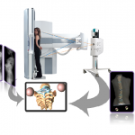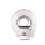
Sponsored • The IQon Spectral
Accustomed workflow, low dose and more precise diagnostics
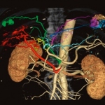
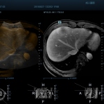
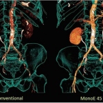
In spectral imaging, x-ray images are formed in the customary grey scale imaging procedure. However different photon energies are used, generating images in different colors. Aside from the acquisition of anatomical information, this measurement makes it possible to show different tissue compositions.