Sponsored • Orthopedics
Spinal imaging
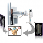
Last year, the DMS Group acquired 100% of the capital of AXS Medical, a French start-up specialized in developing diagnostic tools used for spinal pathologies.

Last year, the DMS Group acquired 100% of the capital of AXS Medical, a French start-up specialized in developing diagnostic tools used for spinal pathologies.

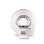
A pioneer in cone beam computed tomography (CBCT) imaging, NewTom recently introduced the only CBCT system with an open gantry and supine positioning, which the firm reports is ‘…ideal for a host of diagnostic needs. Exceeding the limits posed by CT systems, the NewTom 5G XL combines high diagnostic resolution with minimum patient exposure.’

The annual congress of the ESR is a great showcase for the latest advances in the imaging sciences and an important platform to see where the industry is heading.

MRI, CT, pathology: doctors have to consider medical image data – increasing in both amount and complexity – to perform diagnoses and monitor therapy. The Fraunhofer Institute for Medical Image Computing MEVIS in Bremen is creating a new approach to provide effective assistance.

Taking clear pictures of megaenzymes isn’t easy. But it’s definitely worth it. These proteins play an active role in creating many common antibiotics. They are in constant motion, with sections that flip around acrobatically to carry out necessary tasks. Now, for the first time, McGill researchers have been able to take a series of 3D images of a large section from one of these…

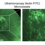
Stopping the growth of blood vessels in tumours is a key target for glioblastoma therapies, and imaging methods are essential for initial diagnosis and monitoring the effects of treatments. While mapping vessels in tumours has proven a challenge, researchers have now developed a combined magnetic resonance imaging (MRI) and ultramicroscopy 'toolkit' to study vessel growth in glioma models in more…

Eckert & Ziegler AG, a specialist for isotope-based applications in medicine, science and industry, is expanding its cooperation program with promising drug developers in the field of nuclear medicine and will support Curasight, a spin-off based on research by the group of Professor Andreas Kjaer at the National University Hospital (Rigshospitalet) and University of Copenhagen, in obtaining…

A new way of using MRI scanners to look for evidence of multiple sclerosis in the brain has been successfully tested by researchers at The University of Nottingham and Nottingham University Hospitals NHS Trust.

Scientists of the Göttingen Cluster of Excellence CNMPB and the EMBL describe efficient fluorescence tag for super-resolution microscopy.

Citing concerns over attendee safety, the European Society of Thoracic Imaging (ESTI) has cancelled its 2016 annual scientific meeting which had been scheduled for May 25 to 28 in Istanbul.

A group of researchers from Russia, Australia and the Netherlands have developed a technology that can reduce Magnetic Resonance Imaging (MRI) scanning times by more than 50 percent, meaning hospitals can drastically increase the number of scans without changing equipment. This extraordinary leap in efficiency is achieved by placing a layer of metamaterials onto the bed of the scanner, which…

Doctors at the Duke University School of Medicine have tested a new injectable agent that causes cancer cells in a tumor to fluoresce, potentially increasing a surgeon’s ability to locate and remove all of a cancerous tumor on the first attempt. The imaging technology was developed through collaboration with scientists at Duke, the Massachusetts Institute of Technology (MIT) and Lumicell Inc.

Breast cancer screening has helped to detect cancer in its early stages, but it is unclear how important this contribution is to mortality reduction because treatment has greatly improved. Overdiagnosis and overtreatment remain associated risks that need to be fully assessed for screening to be of real benefit. Leading experts in this field passionately discussed these controversies in a…

More is better – when it comes to medical display technology a higher resolution is a desirable feature of next generation displays. Today, the number of pixels alone is just one of many factors that distinguishes a display developed specifically for diagnostic purpose from those designed for regular use. Shinji Nohara, Product Manager for Pro/Colour/Medical Desktop Display at NEC Display…

With the aim of producing high-quality X-rays with minimal radiation exposure, particularly in children, researchers have developed a new approach to imaging patients. Surprisingly, the new technology isn’t a high-tech, high-dollar piece of machinery. Rather, it’s based on the Xbox gaming system.

Ziehm Vision RFD 3D is the world’s only 3D C-arm with flat-panel technology that provides a 16 cm edge length per scan volume combines 2D and 3D functionality to offer maximum intraoperative control.

Using the Arietta V70 from Hitachi, a French diagnostic imaging team is rewriting the book on obstetrics and gynaecology. Entitled the ‘Atlas d’échographie de fusion en gynécologie obstétrique’, the new edition by Jean-Marc Levaillant MD, and colleagues from the diagnostic imaging centres at the Bicêtre and Créteil hospitals in Paris, will be published before the end of 2015.

In mid-2015 the second generation of the ultrasound system H60 and the Urostation Touch was launched. At the Urology Department of the Hospital Fürth in Fürth, Germany the H60 and Urostation Touch are already being successfully used with 4 to 5 patients a week for prostate diagnostics.

Impacting on clinical decisions. Accelerating clinical routine. Following the release of its new Version 6 software upgrade for the Aplio Platinum Series ultrasound system, Toshiba has received high marks for the enhanced functions and performance from practitioners, each offering specific insights into how they are applying the technology.

This year at MEDICA, Sony Healthcare Solutions builds on its position in the market as a leading provider of medical imaging technologies. Sony will demonstrate the power and impact visual solutions have in the world of medicine, from pre-operative diagnosis and the latest 4K surgical imaging through 4K over IP distribution up to education, post-surgical review and long archive storage.

Trice Imaging announced today at Health 2.0 the integration of Tricefy into FUJIFILM SonoSite’s latest portable ultrasound device. The iViz, which was recently CE marked for sale in Europe and currently pending FDA 510(k) clearance, is the latest point-of-care visualization solution by FUJIFILM SonoSite and includes Trice sharing, collaboration and routing software embedded in the device. Both…
Large patient volumes and a shrinking pool of skilled resources in the European healthcare industry are creating the need for efficient workflow solutions. Hence, hospitals will increasingly invest in digital radiography (DR) as the revival of the regional economy picks up pace. While the saturated computed radiography (CR) segment is declining in terms of both unit shipment and revenue, the…

Tweets can give medical professionals a window into the minds of patients, according to a new study published in the Journal of Medical Imaging and Radiation Sciences.

Dirk-André Clevert MD, Associate Professor of Radiology at the Department of Clinical Radiology, University of Munich, Germany, describes a new diagnostic approach in abdominal ultrasound.

At Agfa HealthCare, we have long recognized the need for a balance between high image quality and the management of dose. With the cesium phosphor technology enhanced and empowered by the gold standard MUSICA image processing software, renowned for delivering diagnostic value images and making it easy to see details, radiologists are potentially able to provide a dose reduction of 50% to 60%*.…