News • Non-invasive diagnostics
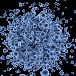
Detecting bladder cancer with atomic force microscopy
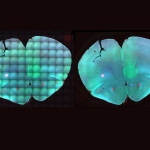
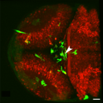
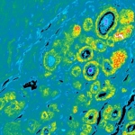
A research team led by Tufts University engineers has developed a non-invasive method for detecting bladder cancer that might make screening easier and more accurate than current invasive clinical tests involving visual inspection of bladder. In the first successful use of atomic force microscopy (AFM) for clinical diagnostic purposes, the researchers have been able to identify signature features…