Improved and faster analysis of MR images
Thanks to a new software, MR images from a five-minute scan may generate the images for a complete and comprehensive examination and therefore eliminating the need to perform multiple scans. The advanced software developed by Synthetic MR AB will be offered as a clinical application in Sectra´s PACS system.
Based on these maps any T1 weighted and T2 weighted image can be synthesized
“We are highly impressed by the efficiency enhancement in magnetic resonance examinations that may potentially be achieved with Synthetic MR´s software,” says Torbjörn Kronander, President of Sectra Imtec AB. The company recently has signed a cooperation agreement with the Swedish research Company Synthetic MR AB with the result that Sectra now can offer the advanced software that provides time-savings for the patients, hospital personnel and technicians as well as facilitating and improving the analysis of MR images.
A normal examination of clinical Magnetic Resonance Imaging (MRI) consists of the generation of a series of contrast images on a patient. The image pixel intensity of these contrast images depends on both patient specific MR tissue parameters, such as the longitudinal T1 relaxation, the transverse T2 relaxation and the proton density (PD), and on the other hand the MRI scanner settings with parameters such as the echo time (TE), the repetition time (TR), the flip angle and the application of preparation pulses. During an examination the scanner settings are predefined to particular values and subsequently the scan is performed. This procedure is repeated over and over again with different settings until all desired contrast images are acquired. This can be a time-consuming procedure, taking 45 - 60 minutes.
A better approach is the direct measurement of the absolute MR tissue parameters for tissue characterization. A quantitative measurement allows to estimate the absolute deviation from the normal and the progress of disease over time. Examples for the clinical relevance of absolute quantification are the use of T1 relaxation time for diseases such as Parkinson, Alzheimer and Multiple Sclerosis, or T2* relaxation time to assess iron deposition in e.g. thalassemia, or the use of several MR parameters for the characterization of atherosclerotic plaques.
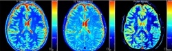
In spite of the clinical benefits of MR tissue quantification its application is far from common clinical practice. The two most important reasons are the associated scan time for accurate quantification and the appearance of the data set. At the Center of Medical Imaging Science and Visualization (www.cmiv.liu.se) a rapid and flexible method for simultaneous quantification of the MR tissue parameters is developed in order to overcome the first hurdle. Instead of the usual multi-hour measurement a complete brain can now be accurately captured at high resolution in about 5 minutes which allows absolute quantification to enter the clinical arena. The second hurdle is addressed using the concept of Synthetic MRI. Based on the absolute MR tissue parameters it is possible to synthesize any T1 or T2 weighted contrast image that is common in clinical practice. This means that the mentioned 5 minutes scan may generate the images for a complete and comprehensive examination. The similarity of the synthetic images with the normal images will significantly lower the threshold for application of absolute quantification in MRI. Moreover, using this approach images can be optimized even if the patient has already left and it is even possible to visualize stronger, non-physical perceptualizations that cannot be generated on the MR scanner itself, e.g. Synthetic Vector MRI or Synthetic Color MRI.
The optimization of contrast images by means of Synthetic Contrast MRI, the visualization of various non-physical contrasts and the visualization of Synthetic Color MRI based on absolute tissue values are under patent. Currently the MR sequences for quantification are in their validation phase. Sites can licence the approach and get support. The expectation is that the MR community will quickly adopt Synthetic MRI as a diagnostic and research tool once a proper validation has been done.
06.01.2008