New technology enhances X-ray images
An innovative method to produce dark-field X-ray images was recently presented by researchers from the Paul Scherrer Institute (PSI) and the EPFL in Switzerland. Dark-field images offer more details than ordinary X-ray radiographs, allowing to diagnose early stage breast cancer, osteoporosis or Alzheimer's disease.
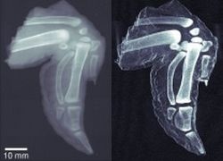
left: traditional X-ray image
right: more detailed dark-field image
© Franz Pfeiffer
Traditional X-ray images show a simple absorption contrast, whereas dark-field images capture the scattering of the radiation within the material itself. The image quality is striking – even subtle inner changes in bone or soft tissue are exposed.
Dark-field X-ray images can be used to detect malignant growth in soft tissue, as cancer cells scatter radiation differently than normal cells. The dark-field technique could also be able to enhance security screening equipment to better identify hidden explosives.
Until recently, dark-field X-ray imaging required sophisticated optics and could only be produced at facilities like the PSI's 300m-diameter, $200 million synchrotron. In their study the researchers describe new nanostructured gratings, that enable ordinary X-Ray equipment to produce dark-field images.
"We have been working on dark-field X-ray images for many years," explains Franz Pfeiffer, professor at the EPFL and researcher at the PSI. "Our new technique uses novel X-ray optical components, in the form of nanostructured gratings, that permit the use of a broad energy spectrum, including the standard range of energies in traditional X-ray equipment used in hospitals or airports," adds Christian David, Pfeiffer's colleague at PSI. "This opens up the possibility for adapting current imaging equipment to include dark-field imaging."
A collaboration is planned with the Center for Biomedical Imaging (CIBM), a joint center with the Universities of Lausanne and Geneva and their associated hospitals. The researchers goal is to develop an adaptation for existing medical equipment. "When combined with the phase contrast imaging technique that we developed in 2006, we now have the possibility of providing the same range of imaging techniques in broad-spectrum X-ray imaging that we do with visible light," Pfeiffer concludes.
For more information please click here (Ecole Polytechnique Fédérale de Lausanne website).
21.01.2008