Mega-MRI
Pushing the power of scanners creates new world for imaging, but whilst high-field magnets bring new capabilities they also pose new challenges for clinicians.
John Brosky reports
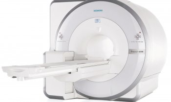
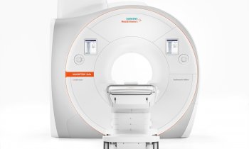
Magnetic resonance imaging (MRI) has become so powerful it needs to be moved to a building of its own on a hospital campus. Currently some 50 new MRI scanners at 7-tesla have been installed worldwide, an impressive increase in power from the top-of-the-line 3-T machines that can be found in advanced radiology departments, and far beyond the everyday MRI of 1.5-T used for routine clinical exams.
‘These are expensive machines, but the scanner is the cheapest part,’ said Professor Jürgen Hennig, of the Department of Radiology at the University Hospital Freiburg. ‘You need to buy real estate to isolate the MRI, and then tons of metal sheeting to insulate the machine. Taking MRI to 7-T is not just performing more of the same examinations, it moves us into a different world,’ he told colleagues during a special session at the ECR in March.
In the New Horizons Session Quantum Leaps in MRI, leading clinicians described the dazzling possibilities for muscular MRI that renders vivid images of the brain, or the heart, or an exquisite precision for the study of new tissue growth in knee cartilage. Yet, despite all this potential, ‘there is not a killer clinical application,’ acknowledged Prof. Hennig.
Pushing up the power of the magnet also creates a new world of complications, primarily for the radio frequency coils that detect the signals, according to Luc Darrasse, Director for MRI at the French National Research Centre.
The heart, the knee and the human brain are objects too small for the enormous magnetic forces requiring a new generation of coils capable of capturing complex signals in parallel, and then next-level processing to filter the data.
Cardiac studies with high field MRI combined with contrast agents for functional imaging are coming along nicely, reported Silvio Aime, head of the Molecular Imaging Centre of the University of Torino.
The development of high-field MRI will come in two phases. First, functional MRI needs to pass a test using positron emission tomography (PET), the most widely used contrast agent in computed tomography (CT).
In 2010 over three million PET-CT scans were performed worldwide, making this technique the gold standard for functional imaging.
The recent introduction of PET-MRI from Philips with its Ingenuity TF, and Siemens with its Biograph mMR, will now enable MRI researchers to validate clinical applications for an alternative method.
‘MRI adds to PET a number of advantages looking at soft tissues, and without any radiation,’ said Prof. Aime, adding: ‘There is a huge advantage with MRI to show areas of perfusion uptake and changes in vascular permeability a widely used method to classify tumours.’
‘I would bet on oncology for early tumour response as an application where MRI will pay off,’ predicted Prof. Hennig, adding: ‘The brain is an exciting application, where we can show micro-bleeding, which CT can not. For Alzheimer's, it’s much clearer with MRI where the tracer is accumulating. Beyond the brain, bones and joints are appropriate to the technology and the new MR centre in Vienna is providing spectacular images.’
In his presentation, Prof. Aime described how MRI can move beyond radioactive PET tracers in the blood with contrast agents custom designed to search for disease in the body.
Chemistry offers a brilliant array of nano carriers loaded with a binding agent that finds a specific condition and an imaging agent that ‘lights up the region of interest’, he said. These carriers can also be loaded with a drug to treat the condition, creating the possibility with MRI to visualise the delivery of a therapy directly to a disease site. ‘This opens new fields for theranostics, therapies combined with diagnostics that will improve the treatment of patients,’ Prof. Aime pointed out.
A self-described ‘MRI guy,’ Prof. Hennig is more enthusiastic about this second phase for high-field MRI where custom-designed MRI probes will illuminate our understanding of disease and treatment. ‘First, of course, we need to validate that MRI can acquire information that is as good or even better than CT,’ he said. ‘Then we can switch off the PET and explore new possibilities.’
20.04.2011


