New MRI methodology revolutionizes imaging of the beating heart
Scientists of the Charité – Universitätsmedizin Berlin and the Max-Delbrück-Center for Molecular Medicine (MDC) Berlin-Buch have developed a highly efficient approach for imaging the beating human heart. The images produced in one of the world's most powerful MRI systems whose power is equivalent to 150.000 times the earth’s magnetic field are of a much higher detail than cardiac images commonly generated in current clinical practice.
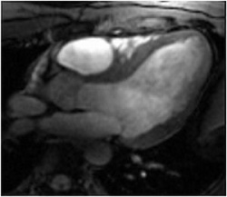
The ultrahigh field approach permits a superb delineation between blood and heart muscle. Even subtle anatomical structures are made clearly visible. The new procedure holds the promise to advance the capabilities of cardiac research and care as cardiac malfunctions can be diagnosed, treated and monitored at a much earlier point in disease progression.(1)
For cardiac imaging in ultrahigh fields new versions of multi-channel transmit and receive antennas - so-called radiofrequency coils - were developed at the Berlin Ultrahigh Field Facility (B.U.F.F.) located at Campus Buch. For this purpose a joint collaboration between the Charité, the MDC, the German Metrology Institute and Siemens Healthcare was initiated.(2) To make use of the capacity and traits of the strong magnetic field a groundbreaking triggering device was developed to synchronize cardiac imaging with heart motion.(3) This approach eliminates mis-synchronization frequently encountered with conventional triggering devices and hence helps to generate crisp cardiac images, a feature which might be compared with sport macros used in digital photography. "We correlate the image exposure with the heartbeat" explains the investigator of the study Prof. Thoralf Niendorf, whose work is published in the March issue of the journal for Magnetic Resonance Imaging.2 "Our procedure is immune to interference with strong magnetic fields so that we can compensate for the motion of the heart which results in high image quality free of cardiac motion induced blurring and artifacts".
The Berlin-based team led by Professor Thoralf Niendorf, Prof. Jeanette Schulz-Menger from the Charité and Dr. Bernd Ittermann from the German Metrology Institute used the new technologies to derive for the first time a clearly defined image of the beating heart in a magnetic field with a strength of 7.0 Tesla. The advancement in imaging technology culminated in images of the beating heart with a spatial resolution which is by far superior to that previously available and which might come close to turning a 10 megapixel digital camera into a 50 megapixel digital camera. The novel technology tailored for cardiac MRI together with the quality of the anatomical and functional images have created excitement among the international imaging community. The first clinical results and experiences were very much encouragingand are the driving force for broader clinical studies.(4)
1. von Knobelsdorff-Brenkenhoff et al.: Cardiac Chamber quantification using magnetic resonance imaging at 7 Tesla--a pilot study. Eur Radiol. 2010 Dec;20(12):2844-52. Epub 2010 Jul 17. doi: 10.1007/s00330-010-1888-2.
2. Dieringer et al.: Design and application of a four-channel transmit/receive surface coil for functional cardiac imaging at 7T. Journal of Magnetic Resonance Imaging, 2011 March, 33(3):736-41. doi: 10.1002/jmri.22451. Epub 2011 Feb 1.
3. Frauenrath et al.: Acoustic cardiac triggering: a practical solution for synchronization and gating of cardiovascular magnetic resonance at 7 Tesla. J Cardiovasc Magn Reson., 2010 Nov 16;12:67. doi: 10.1186/1532-429X-12-67.
4. Niendorf et al.: Toward cardiovascular MRI at 7 T: clinical needs, technical solutions and research promises. Eur Radiol. 2010 Dec;20(12):2806-16. Epub 2010 Jul 31. doi: 10.1007/s00330-010-1902-8.
27.04.2011