
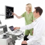
Cause for concern: Diagnoses via teleradiology
Whilst the benefits of teleradiology as a diagnostic tool continue to grow, concerns have been raised about the lack of uniformity in reporting protocols across Europe. Mark Nicholls reports


Whilst the benefits of teleradiology as a diagnostic tool continue to grow, concerns have been raised about the lack of uniformity in reporting protocols across Europe. Mark Nicholls reports
Carestream Health provides innovative digital radiography systems that satisfy these goals while simultaneously helping to reduce expenses for healthcare providers worldwide. It also markets powerful systems that efficiently manage radiology workflow—from the scheduling of patient x-ray exams to reading of those imaging studies by radiologists, and ultimately the delivery of radiology reports…
Royal Philips Electronics is announcing 510(k) clearance from the Food and Drug Administration (FDA) for the company’s first commercially available whole body positron emission tomography/magnetic resonance (PET/MR) imaging system, the Ingenuity TF PET/MR.
By combining conventional medical imaging with some of the same 3-D modeling techniques used in Hollywood blockbusters, researchers are offering new hope to victims of serious facial injuries. Results of a new study on human face transplantation, led by Darren M. Smith, M.D., plastic surgery resident at the University of Pittsburgh Medical Center (UPMC), were presented today at the annual meeting…
Agfa HealthCare announces that it will launch its automated DX-D 600 direct radiography (DR) system, which has received 510k clearance by the U.S. FDA, into the U.S. market at RSNA 2011. Combining user-friendly design with excellent image quality in a high-productivity direct digital X-ray room, the fully automatic system also offers the latest in state-of-the-art auto-positioning technology.
Amid increased scrutiny over medical imaging scans and the use of radiation, a new survey reveals that awareness and familiarity with medical imaging tests lead to clearer decisions for U.S. adults about their healthcare. The survey, released by the Siemens Radiation Reduction Alliance (SIERRA) – an expert panel established to advance the cause of dose reduction in medical imaging – evaluated…
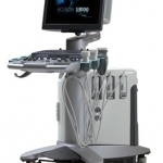
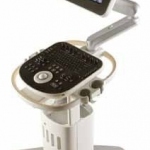
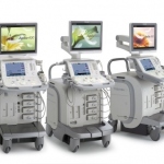
Siemens Healthcare launched the ACUSON S3000, its latest ultra-premium ultrasound platform, at the 97th Scientific Assembly and Annual Meeting of the Radiological Society of North America (RSNA) in Chicago, USA. The new system includes advanced automated ultrasound fusion imaging as well as multi-modality review capabilities to provide additional clinical and spatial information in the analysis…

Portable ultrasound just got more compact with the first transducer embedded with a full scanning system that plugs into the USB port of tablet or laptop computers.
Based in Guangdong, China (VR), the Shantou Institute of Ultrasonic Instruments Co. Ltd. (SIUI) has been engaged in the development and manufacture of medical ultrasonic diagnosis scanners, industrial ultrasonic detectors and ultrasonic transducers since 1978. The company reports that, from its beginning as a small scientific research institute, through the years SIUI has developed into a key…
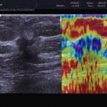
Dr Katja Gabriel of the Hirschhauser & Gabriel Obstetrics and Gynaecology Practice in Erkrath, Germany, describes clinical experience with this novel technique and the resulting improved diagnostic accuracy.

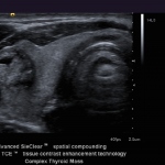
In August, Bill Smith, Head of Ultrasound Services at privately-owned Clinical Diagnostic Services in London,UK, was particularly excited about advances in elastography and Fly Thru in Toshiba’s newly launched high-end ultrasound series Aplio 300, 400 and 500. Discussing initial experiences utilising the system, he said, ‘Fly Thru is opening up completely new perspectives for noninvasive…

With most radiology departments using digital equipment, smoothing image and data mnagement, CEOs and medical heads of departments aim to achieve those benefits – and particularly paperless systems – across their hospitals. HCI, a consulting and implementation company based in Brilon, Germany, specialises in integrating systems in, for example, operating theatres. Formerly employed in key…
Manufactured by Royal Philips Electronics and currently being introduced in Europe, India, Australia and New Zealand, ClearVue introduces proprietary Active Array technology, which moves key technology from the system to the transducer, resulting in enhanced image quality in 2-D and colour, lighter weight cabling and increased transducer reliability, Philips reports.

The goals are ambitious: Although in the market merely four years, the start-up firm Alpinion Medical Systems states its intention to become one of the prime providers in the ultrasound segment with superior imaging and unique transducer technologies. In an interview with Daniela Zimmermann of European Hospital, Thomas Roth, Alpinion’s Managing Director, explains his corporate strategy and…
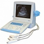
The Honda HS-2100 is one of the latest developments from the Medical division of Honda Electronics, in Aichi, Japan. This portable, sleek, compact system has fine image quality, the firm reports: The front-end is fully digital which, combined with the unique technology, guarantees optimal image quality and focusing on pixel level.
Siemens Healthcare reports that the firm’s new Acuson S2000 system includes the new multifunctional transducer 6C1 HD (high density) specifically designed for abdominal examinations, displaying even the smallest lesions at greater depths. At Medica, the company is also demonstrating its Virtual Touch Tissue Analytics technology, a proprietary implementation of Acoustic Radiation Force Impulse…
SonoAce GmbH, the subsidiary of Samsung Medison based in Germany, has announced the development of two ‘outstanding new imaging technologies’
Toshiba’s new high-end ultrasound series, the Aplio 300, 400 and 500, introduced during the World Congress of Ultrasound in Medicine and Biology in Vienna, this August, was heralded by the firm as ‘A giant leap forward’, due to its inclusion of features such as Fly Thru and Smart Fusion.

Siemens Healthcare is the first medical engineering company to offer a Low Dose Information Center on the Web. This English-language platform for continuing education and information around the topic of dose reduction is aimed at doctors and clinical personnel. The range of topics covered includes basic information on X-ray radiation, technical innovations relating to dose reduction and sample…

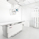
Ziehm Imaging’s C-arm is a general X-ray unit. It’s what hospitals really need, says Timo Ihamäki, the firm’s business manager, because ‘It’s a mobile unit that can be moved to the OR, out of the OR, to the trauma room, A & E, or wherever needed -- it’s always in the hallway. Whoever needs it, takes it.’

In an interview with Daniela Zimmermann, Professor Jerzy Walecki explained the implications of that role and the state of radiology in Poland today.
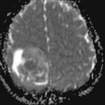
As a referral neuroradiologist for paediatric tumour studies, Professor Monika Warmuth-Metz, Consultant at the Neuroradiology Department at University Hospital Würzburg, daily evaluates MRI images of different origin and colour. Her resume states: ‘All too often the standard protocols set out in the guidelines are not adhered to, which makes evaluation and follow-up significantly more…
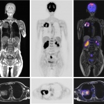
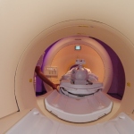
Although like a conventional MR scanner the unassuming exterior is misleading. The casing houses a powerful interior. This is the new Siemens Biograph mMR, a hybrid that contains a specially developed PET component fully protected against magnetic field interference.
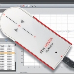
The Nomex multimeter, a miniaturised non-invasive measuring system for absolute dosimetry and quality control in X-ray diagnostic radiology, will be demonstrated in several upcoming European medical trade shows*, including Medica in Dusseldorf.
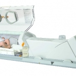
A solution for a large but previously unsolved problem has been solved by LMT Lammers Medical Technology GmbH, which specialises in the interface of neonatology and radiology.