From pre-natal diagnostics to 3-D baby-facing
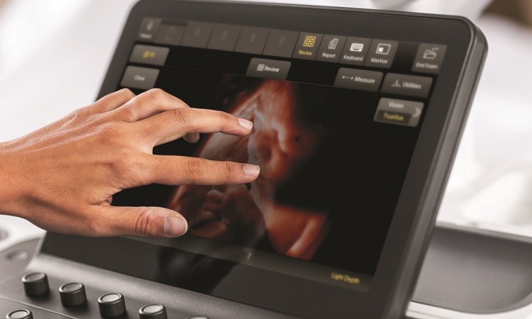
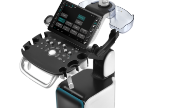
Prenatal ultrasound images are the first images we see of new humans. But the prenatal ultrasound first carried out by Professor Ian Donald in the late 50s has little to do with today’s potential with 3-D and 4-D imaging with Doppler and colour Doppler. The three tightly linked disciplines gynaecology, prenatal medicine and obstetrics would now be inconceivable without ultrasound
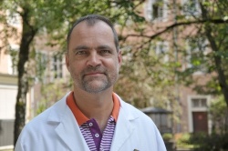
Inevitably, gynaecology and obstetrics will be key WFUMB topics this year, with pre-natal ultrasound the most important subject, says Professor Horst Steiner, Assistant Director of the University Clinic for Gynaecology and Obstetrics at the Paracelsus Medical Private University in Salzburg, Austria, and member of the WFUMB gynaecology, prenatal medicine and obstetrics sub-committee.
‘The most important innovation is that we can now answer many questions around the health of the unborn child in the first trimester, i.e. between the 11th and 14th week of pregnancy,’ he explains. ‘This includes anatomical development. The most common morphological changes in the foetus are congenital heart defects, which occur in 1% of cases. Ultrasound can also be used to check genetic defects. The detection rate for trisomies, such as Down's Syndrome, in combination with blood tests is above 90%.’
Risk examinations are carried out, for example, in patients who have suffered complications in previous pregnancies. The average age for pregnancy is on the increase and nowadays multiple births are by no means rare, so gynaecologists are increasingly being faced with high risk pregnancies.
A large part of prenatal ultrasound is involves screening. Apart from standard examinations, now there are many highly-complex additional services on offer. These can be utilised if medically indicated or simply desired by the patient. Many mothers- to-be are prepared to pay for such enhanced screenings without any medical need for them, Prof. Steiner points out. ‘They just want to know as early as possible whether there is likely to be a serious problem or not. The important thing is to make them aware of the possible consequences of such examination, such as changes in pregnancy care, or the hospital chosen for the birth right down to situations where there may indeed be a reason to terminate a pregnancy.’
Prof. Steiner sees a need for a clear distinction between prenatal diagnostics to detect problems and deformities and the so-called ‘baby-facing’. Many pregnant mothers simply want to see their unborn children without there being any medical indication for these scans. This is understandable and in many cases a good thing -- for example to put at rest the mind of a patient who has experienced a previous, problematic pregnancy. There are also studies that document that the emotional tie between a mother and her unborn child is enhanced through so-called baby-facing. It’s important to make parents-to-be aware of the difference between medical diagnostics and ‘baby-facing’.
Obstetric ultrasound and prenatal diagnostics are other important topics at the event.
As equipment is increasingly sized down, ultrasound is also being used in the delivery room. ‘Basic examinations, such as ascertaining the position of the baby in the birth canal, can be conveniently carried out on site,’ Prof. Steiner points out. Doppler ultrasound can be used to monitor the haemodynamics of the child and the placenta to see if there are any problems. We are increasingly able to monitor the birth mechanics trans-abdominally and trans-labials and can intervene when problems occur.’
Gynaecological ultrasound, including for non-pregnant patients will be another focus at the congress. Apart from breast diagnostics, the classic gynaecological applications are for malignoma treatments.
‘Clinical investigation of elastography is quite a recent development in gynaecology. There are first experiences in this field, but unfortunately no groundbreaking results as yet. Moreover, the use of contrast media could bring new momentum to the diagnosis of ovarian tumours,’ Prof. Steiner believes. ‘But it’s as yet completely unclear whether ultrasound contrast media will result in a better prognosis with regards to dignity determination.’
Lastly the professor sees much potential in emergency ultrasound. ‘This is an interdisciplinary field that also covers numerous gynaecological problems, such as ectopic pregnancies, bleeding cysts or bleeding tumours. Ultrasound makes a unique diagnostic contribution in emergencies due to its high mobility and fast availability.’
"US in the delivery room/Doppler techniques", Saturday, August 27, 16:00-17:30, Hall D
Horst Steiner
Professor Horst Steiner, Assistant Director and head of out-patient services for prenatal medicine at the University Clinic for Gynaecology and Obstetrics in Salzburg, Austria, gained his medical doctorate in Innsbruck in 1983 and completed his specialist training in gynaecology and obstetrics at the Women's Clinic in Salzburg, specialising in urology, general surgery and neonatology. Residencies followed in the USA and Sweden.
Prof. Steiner, who is also a Board Member of the Austrian Society of Gynaecology and Obstetrics, regularly holds training courses in Doppler sonography and ultrasound diagnostics, has given more than 300 lectures and is author and co-author of some 200 publications.
25.08.2011


